[level-membership-for-gastroenterology-and-hepatology-category]CASE 147
History: A 41-year-old Malaysian man presents with fever.
1. What should be included in the differential diagnosis of the imaging finding showing an abnormal cecum in Figure A? (Choose all that apply.)
2. What is the most common site of involvement of abdominal tuberculosis?
3. Which statement regarding tuberculosis of the gastrointestinal tract is true?
4. Several other infections favor the ileocecal region; which of the following infections does not?
ANSWERS
CASE 147
Tuberculosis of the Cecum
1. A, B, and C
2. B
3. D
4. A
References
Burrill J, Williams CJ, Bain G, et al: Tuberculosis: a radiologic review. Radiographics. 2007;27(5):1255–1273.
Leder RA, Low VH. Tuberculosis of the abdomen. Radiol Clin North Am. 1995;33(4):691–705.
Cross-Reference
Gastrointestinal Imaging: THE REQUISITES, 3rd ed, p 300.
Comment
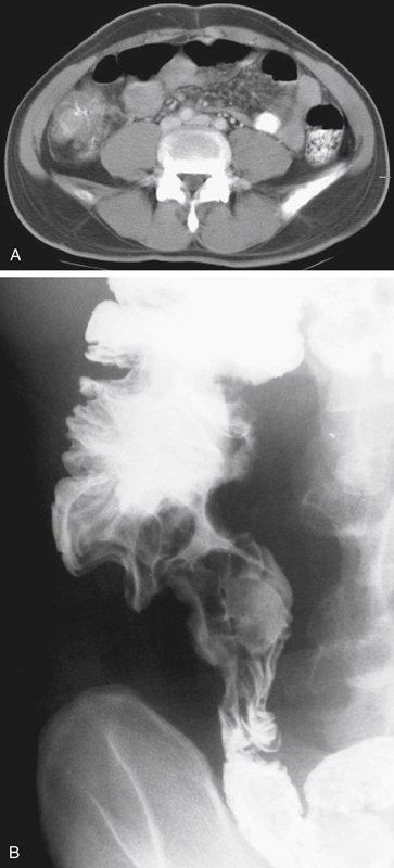
The CT image shows thickening and deformity of the cecum with a narrowed lumen (see figures). The image from the barium small bowel follow-through in this case shows some deformity of the cecum (see figures). One of the most common inflammatory processes of the ileocecal region is Crohn’s disease. It also can affect any portion of the gastrointestinal tract, and the colon is a likely area for further involvement. Possible infectious processes include amebiasis and tuberculosis. Both of these conditions can affect the ileocecal area and the rest of the colon. Of the possible neoplastic processes, the most likely is serosal metastasis, which has an affinity for the cecal region. Lymphoma must always be considered in the differential diagnosis of gastrointestinal lesions.
The final correct diagnosis for this patient was tuberculosis. When a chest radiograph was obtained, it was normal. A normal chest x-ray film should not preclude this diagnosis because intestinal tuberculosis can occur without an abnormal chest film, especially in patients with human immunodeficiency virus. Most intestinal tuberculosis occurs as a result of a patient with pulmonary tuberculosis swallowing infected sputum; intestinal tuberculosis is usually secondary. Although adenopathy may occur in patients with intestinal tuberculosis, it also occurs in patients with Crohn’s disease or lymphoma and does not help differentiate the conditions. Patients with these findings in the right lower quadrant are often misdiagnosed as having Crohn’s disease.
[/level-membership-for-gastroenterology-and-hepatology-category][not-level-membership-for-gastroenterology-and-hepatology-category]CASE 147
History: A 41-year-old Malaysian man presents with fever.
1. What should be included in the differential diagnosis of the imaging finding showing an abnormal cecum in Figure A? (Choose all that apply.)
2. What is the most common site of involvement of abdominal tuberculosis?
3. Which statement regarding tuberculosis of the gastrointestinal tract is true?
[/not-level-membership-for-gastroenterology-and-hepatology-category]
