CASE 144
1. What features are characteristic of a malignant cardiac tumor? (Choose all that apply.)
B. Involvement of two or more chambers
C. Extension outside of the heart
2. What is the most common tumor in the heart?
A. Fibroma
B. Myxoma
C. Metastasis
D. Hemangioma
A. Left atrium
C. Right atrium
4. What is the most likely diagnosis?
A. Hemangioma
B. Lipoma
C. Myxoma
D. Fibroma
ANSWERS
References
Moniotte S, Geva T, Perez-Atayde A, et al. Images in cardiovascular medicine Cardiac hemangioma. Circulation. 2005;112(8):E103–E104.
Randhawa K, Ganeshan A, Hoey ET. Magnetic resonance imaging of cardiac tumors: part 1, sequences, protocols, and benign tumors. Curr Probl Diagn Radiol. 2011;40(4):158–168.
Cross-Reference
Cardiac Imaging: The REQUISITES, ed 3, p 280.
Comment
Histology and Physiology
Hemangiomas are benign proliferations of endothelial cells and vessels. These masses can be classified as a cavernous, a capillary, or an arteriovenous subtype, depending on the dominant vascular channel. Hemangiomas can contain calcification, fat, and fibrous tissue. Cardiac hemangiomas are rare and can be intramural or within a chamber.
Clinical symptoms and signs include dyspnea on exertion, arrhythmia, angina, and right heart failure. Right ventricular outflow obstruction is the usual cause of dyspnea in patients with hemangioma.
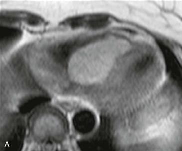
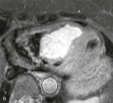
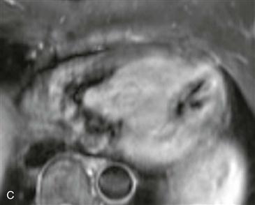
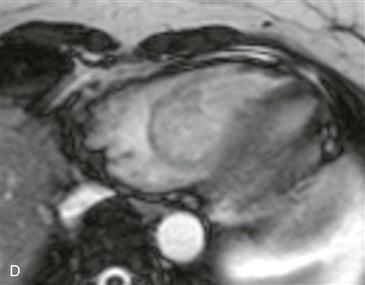
CT and MRI
CT can be used to delineate the location, size, and extent of the mass. On CT, a hemangioma is typically heterogeneous and often demonstrates areas of calcification. Iodinated contrast agent is useful to demonstrate marked enhancement of this vascular mass. MRI can provide optimal evaluation of a cardiac mass (Figs. A–D). Hemangiomas demonstrate low to intermediate signal intensity on T1-weighted images and high signal intensity on T2-weighted images, but occasionally the mass also shows high signal intensity on T1-weighted images. When a mass demonstrates high signal intensity on T1-weighted images, it is important to consider the diagnoses of lipoma and melanoma in addition to hemangioma. Hemangiomas do not lose signal with fat suppression techniques, allowing differentiation from lipomas. Melanoma metastases and other malignant tumors are infiltrative, in contrast to a hemangioma.


