CASE 143
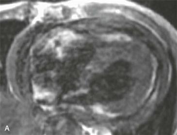
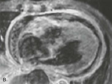
1. What pericardial findings are seen? (Choose all that apply.)
A. Effusion
B. Thickening
D. Enhancement
2. What is the most likely etiology?
D. Tuberculosis
3. Given the symptoms and MRI images, what is the most likely diagnosis?
B. Tamponade
C. Effusive constrictive pericarditis
D. Tumor
4. Which of the following is NOT an MRI imaging finding seen in constrictive pericarditis?
A. Pericardial thickening greater than 4 mm
ANSWERS
References
Syed FF, Ntsekhe M, Mayosi BM, et al. Effusive-constrictive pericarditis. Heart Fail Rev 2012;.
Wang ZJ, Reddy GP, Gotway MB, et al. CT and MR imaging of pericardial disease. Radiographics. 2003;23(Spec No):S167–S180.
Cross-Reference
Cardiac Imaging: The REQUISITES, ed 3, p 269.
Comment
Etiology and Characteristic Features
Effusive constrictive pericarditis is most commonly caused by tuberculosis. Pericardial effusions and pericardial thickening are characteristic. In the setting of constrictive/restrictive physiology, the diagnosis is effusive constrictive pericarditis. Pericardiocentesis can relieve the acute illness and can aid in diagnosis, but chronic constrictive pericarditis can develop.


