CASE 142

History: A patient has hypertension.
1. How can the pressure gradient be measured in coarctation of the aorta? (Choose all that apply.)
B. CT
C. MRI
D. Angiography
2. Which method can be used to measure collateral blood flow in coarctation of the aorta?
A. Transthoracic echocardiography
B. Transesophageal echocardiography
C. CT
D. MRI
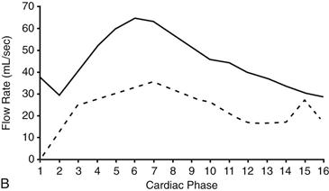
3. What does the graph (Fig. B) represent?
A. Peak velocity across the coarctation
D. Pulmonary-to-systemic flow ratio
4. In a young adult, what is the most appropriate management?
A. No treatment
B. Antihypertensive medication
D. Surgery
ANSWERS
Reference
Hom JJ, Ordovas K, Reddy GP. Velocity-encoded cine MR imaging in aortic coarctation: functional assessment of hemodynamic events. Radiographics. 2008;28(2):407–416.
Cross-Reference
Cardiac Imaging: The REQUISITES, ed 3, pp 72, 420.
Comment
Clinical Features
Congenital coarctation is most commonly discrete and juxtaductal in location (Fig. A). Hypertension is a common finding.
Measurement of Collateral Circulation
Velocity-encoded cine MRI is the only noninvasive method that can accurately quantify collateral circulation in coarctation of the aorta (Fig. B). In normal individuals, flow in the distal thoracic aorta is slightly lower than in the proximal descending aorta because the intercostal arteries and other aortic branches take blood away from the aorta. However, in a patient with a functionally significant coarctation, collateral vessels bring blood into the descending thoracic aorta, and the distal flow is greater than the proximal flow. The presence of collateral flow shows that the lesion is hemodynamically significant, even in the absence of visible collateral vessels.


