CASE 140
1. What should be included in the differential diagnosis based on the history? (Choose all that apply.)
B. Ventricular septal defect (VSD)
D. Transposition of the great arteries
B. VSD
D. Transposition of the great arteries
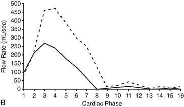
3. What does the graph (Fig. B) represent?
D. Pulmonary-to-systemic flow ratio
4. What is the interpretation of greater pulmonary blood flow (dashed line) compared with systemic flow (solid line)?
ANSWERS
References
Debl K, Djavidani B, Buchner S, et al. Quantification of left-to-right shunting in adult congenital heart disease: phase-contrast cine MRI compared with invasive oximetry. Br J Radiol. 2009;82(977):386–391.
Varaprasathan GA, Araoz PA, Higgins CB, et al. Quantification of flow dynamics in congenital heart disease: applications of velocity-encoded cine MR imaging. Radiographics. 2002;22(4):895–905. discussion 905–906.
Wang ZJ, Reddy GP, Gotway MB, et al. Cardiovascular shunts: MR imaging evaluation. Radiographics. 2003;23(Spec No):S181–S194.
Cross-Reference
Cardiac Imaging: The REQUISITES, ed 3, pp 72, 340–342.
Comment
Echocardiography
Echocardiography is the mainstay of imaging evaluation of cardiac shunts. Echocardiography can be used to depict the anatomy and assess the degree of shunting.
MRI
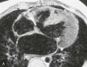
MRI has a key role in the assessment of lesions such as supracristal VSD and partial anomalous pulmonary venous return, for which echocardiographic evaluation may be limited (Fig. A). MRI also has an important role in shunt quantification. Velocity-encoded cine MRI can be used to quantify intracardiac shunts with a great degree of accuracy and reproducibility. Velocity-encoded cine MRI is used to measure blood flow in the pulmonary artery (pulmonary flow) and in the ascending aorta (systemic flow). Flow curves are obtained (Fig. B), and the curves are integrated to obtain flow per unit time. In the normal individual, the pulmonary-to-systemic flow ratio is 1 : 1. However, in a patient with a left-to-right shunt, the ratio is greater than 1. Operative repair may be beneficial when the flow ratio exceeds 1.7:1.


