section 14 Rheumatology and Musculoskeletal
14.1 Rheumatological emergencies
Introduction
The most common rheumatological emergency seen in the emergency department (ED) is acute monoarthritis (see Ch. 14.2). This chapter discusses the important emergencies associated with general rheumatological conditions. Many of these are multisystem diseases and emergencies may be related either to a primary joint problem or to an extra-articular manifestation of the disease. As many of these conditions are autoimmune, suppression of the immune system is usually central to their management. This can be complicated by infection, which can relate to usual pathogens but also opportunistic infection.
RHEUMATOID ARTHRITIS
Management typically involves symptom relief with non-steroidal anti-inflammatory drugs (NSAIDs) and/or corticosteroids, and prevention of disease progression with disease modifying antirheumatic drugs (DMARDs). These include methotrexate, leflunomide, sulfasalazine and hydroxychloroquine. Newer, so-called biologic agents act by inhibiting tumour necrosis factor-α (TNF-α) (infliximab, etanercept, adalimumab) or interleukin-1 (anakinra) and are currently used for patients who fail traditional DMARD therapy. Rituximab is also recently available for those who fail the aforementioned medications – its mechanism of action is B-cell depletion. See Chapter 14.3 for further details on the clinical features of RA, its diagnosis, useful investigations and principles of management.
EMERGENCIES IN RA – ARTICULAR MANIFESTATIONS
Acute monoarthritis
The patient with established RA may present with an acutely painful, hot, swollen joint that may be a manifestation of the underlying condition. Alternatively, it may signal septic arthritis, a condition to which patients with RA are 2–3 times more susceptible than matched controls.1 The possibility of septic arthritis should be considered in a patient with RA who has acute monoarthritis out of keeping with their disease activity. See Chapter 14.2 for the approach to acute monoarthritis.
Cervical spine involvement
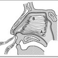
Cervical spine involvement in RA is a common finding with a prevalence of up to 61%.2 It is more common in those with long-standing, erosive disease and disease of greater severity and activity.3 Cervical spine involvement is associated with increased mortality.4 Involvement of the cervical spine may manifest as atlanto-axial subluxation (most commonly anterior movement on the axis) or subluxation of lower cervical vertebrae. Either of these can result in cervical myelopathy.
Cervical spine subluxation is frequently asymptomatic, up to 44% in one study.3 The most common symptom of cervical spine involvement is neck pain that may radiate towards the occiput. Other suggestive symptoms include slowly progressive spastic quadriparesis, sensory loss in hands or feet and paraesthesiae or weakness in the distribution of cervical nerve roots.
Important ‘red flag’ features suggestive of cervical myelopathy are shown in Table 14.1.1.
EMERGENCIES IN RA – EXTRA-ARTICULAR MANIFESTATIONS
Clinical features
Presentations with rheumatoid vasculitis are varied and non-specific. Patients frequently present with constitutional symptoms and fatigue. The most common manifestation is of cutaneous vasculitis with deep skin ulcers on the lower limbs,5 digital ischaemia and gangrene (medium vessels) or palpable purpura (small vessels). Mononeuritis multiplex is another common presentation and results from infarction of the vasa nervorum due to vasculitis. It typically has an acute onset.
Summary of other extra-articular manifestations of RA
Cardiovascular disease in RA and other connective tissue diseases
Patients with RA and other connective tissue diseases such as systemic lupus erythematosus (SLE) have an increased risk of ischaemic heart disease (IHD).6 This occurs independently of traditional risk factors such as smoking or dyslipidaemia, and is more common in those with extra-articular disease.7 The higher incidence of IHD could be related to disease factors such as widespread inflammation, or to medications used such as NSAIDs (including selective COX-2 inhibitors) and corticosteroids.
Assessing SLE disease activity
It is important in the ED to determine SLE disease activity. Useful symptoms of disease activity include mouth ulcers, alopecia and constitutional symptoms, as well as organ-specific symptoms such as arthralgia or pleuritic chest pain.
GIANT CELL (TEMPORAL) ARTERITIS AND OTHER VASCULITIDES
Epidemiology
GCA and PMR rarely occur before the age of 50 years of age.8 The mean age at diagnosis is approximately 72 years, with an incidence of GCA of roughly 1 in 500 of people over the age of 50 years. The incidence and prevalence of PMR are less well studied.
Clinical features
The most common symptom of GCA is headache, usually localizing to the temporal region, although it can be more diffuse. The area is often tender and worsened by brushing the hair. Most patients complain of constitutional symptoms such as malaise, fatigue, anorexia and weight loss. Jaw claudication (pain after a period of chewing) is the most specific symptom for GCA, although not sensitive, as it is only present in 34%.8 On examination the temporal arteries may be thickened, ‘ropey’ and tender with an absent or reduced pulse.
Polymyalgia rheumatica
PMR usually affects the neck, shoulder and pelvic girdles resulting in stiffness and inflammatory pain, worse in the morning and after rest. The pain is often poorly localized and there may be muscle atrophy late in the disease. There can also be evidence of synovitis affecting the shoulders, knees, wrists and hands.
Criteria for diagnosis
The ACR classification criteria for GCA are helpful in differentiating GCA from other forms of vasculitis.9 They include age at onset >50 years, a new headache, temporal artery tenderness or decreased pulsation and an ESR >50. An abnormal artery biopsy showing vasculitis with mononuclear infiltrate or granulomatous inflammation with multi-nucleated giant cells is also required to confirm the diagnosis.
Management
Corticosteroids are the treatment of choice and should not be withheld to perform a biopsy, if there is a strong clinical suspicion. The initial dose for GCA is unclear, but prednisone 1mg/kg/day is usually indicated10 especially for ischaemic complications; however, lower doses such as prednisone 40–50mg have been used.11 The dose of prednisone for PMR alone is lower at 10–20mg/day.11 Most patients do not require hospital admission, provided a temporal artery biopsy can be organized within a few days. However, patients with visual loss at diagnosis require urgent treatment, often with pulsed parenteral corticosteroids, and inpatient admission. Patients with GCA should also be commenced on aspirin.
An approach to the systemic vasculitides
The systemic vasculitides are a group of disorders characterized by an inflammatory infiltrate in the walls of blood vessels resulting in damage to the vessel wall. The clinical manifestations depend upon the size of vessel and location in the vascular tree and may result in systemic or organ-specific manifestations. Table 14.1.2 classifies vasculitic syndromes according to vessel size (there is much overlap).
Table 14.1.2 Classification of systemic vasculitis according to vessel size
| Vessel size | Vasculitis |
|---|---|
| Large | Takayasu’s arteritis |
| Giant cell arteritis | |
| Medium | Polyarteritis nodosa |
| Kawasaki disease | |
| Small | Wegener’s granulomatosis (ANCA+) |
| Microscopic polyangiitis (ANCA+) | |
| Churg–Strauss syndrome | |
| Henoch–Schonlein purpura | |
| Cryoglobulinaemic vasculitis | |
| Leukocytoclastic cutaneous vasculitis |
Differential diagnosis of systemic vasculitis
Management of systemic vasculitis
Treatment is usually with high-dose corticosteroids and, depending on the condition, additional immunosuppression such as cyclophosphamide. Urgent specialist referral is essential.
Emergencies associated with rheumatology therapy
Non-steroidal anti-inflammatory drugs
NSAIDs are used for symptom relief in a variety of conditions, especially inflammatory joint pain. They are of equal efficacy, although those with shorter half lives appear to have less gastrointestinal toxicity.12 NSAIDs should be used in the lowest possible dose for the shortest duration and combination of NSAIDs (not aspirin) should be avoided.12
Corticosteroids
Immunosuppressants/disease modifying antirheumatic drugs
This heterogeneous group of medications is used to prevent joint destruction in the inflammatory arthritides and as steroid-sparing therapy in many connective tissue diseases. They include methotrexate, leflunomide, hydroxychloroquine, sulfasalazine, ciclosporin, azathioprine and cyclophosphamide. Each drug has its own range of adverse effects, but common adverse effects presenting at an ED include cytopenias, rashes including the Stevens–Johnson syndrome, abnormal liver function tests, GI toxicity and heightened susceptibility to infections (see Table 14.1.3).
Table 14.1.3 Adverse effects of disease modifying antirheumatic drugs (DMARDs)
| DMARD | Adverse effects |
|---|---|
| Methotrexate | Nausea and other GI upset, mouth ulcers, abnormal liver function (transaminases), bone marrow suppression, rash, alopecia, pneumonitis Teratogenic Increased bone marrow toxicity in renal impairment – withhold in acute renal failure |
| Leflunomide | Abnormal liver function (transaminases), diarrhoea, rash, alopecia, hypertension, peripheral neuropathy Teratogenic |
| Hydroxychloroquine | Nausea, rash, dizziness (‘cinchonism’), retinal toxicity at higher doses (all uncommon) |
| Sulfasalazine | GI upset, uncommonly abnormal liver function and bone marrow suppression, rashes (rarely, Stevens–Johnson syndrome) |
| Ciclosporin | Renal impairment, hypertension, electrolyte disturbance, hyperuricaemia and gout, gingival hyperplasia, hirsutism |
| Cyclophosphamide | Bone marrow suppression especially neutropenia, GI upset, bladder toxicity, including haemorrhagic cystitis (acute) and bladder cancer (chronic), opportunistic infections Teratogenic |
| Azathioprine | GI upset, rash, systemic symptoms, abnormal liver function, bone marrow suppression, skin cancers, infections |
GI, gastrointestinal.
Presentations of treatment-related emergencies
Bone marrow suppression
Anaemia, leukopenia and thrombocytopenia all occur in patients taking DMARDs such as methotrexate, cyclophosphamide, sulfasalazine and azathioprine. The patient who presents with sepsis and leukopenia is a medical emergency and requires resuscitation, supportive care and broad-spectrum parenteral antibiotics.
Infections
Some studies have reported an increased risk of infection and serious infection in patients receiving anti-TNF therapy, although the largest study of infection risk in patients on anti-TNF agents showed that while there is an increased risk of skin and soft tissue injections, there was no significant difference in risk of serious infections.13 Patients on anti-TNF therapy who develop infections are advised to temporarily cease their treatment and to commence antibiotics. If in doubt, they should be admitted to hospital to receive parenteral antibiotics. There is also an increased risk of re-activation of tuberculosis and possibly of infections with other organisms such as Listeria and Salmonella.13 Rigorous tuberculosis screening prior to commencement of anti-TNF therapy should now be universal.
DMARD-related pneumonitis
Methotrexate and leflunomide both result in lung toxicity. The incidence of methotrexate-induced lung toxicity is difficult to assess but uncommon. The most common type of toxicity is a hypersensitivity pneumonitis, but other forms of lung injury may also occur. Clinical features are non-specific and include constitutional symptoms, cough and progressive dyspnoea. Subacute presentations are more common, although acute and chronic presentations may also occur, with rapid progress to respiratory failure in more acute situations. Patients at higher risk for methotrexate-induced lung injury have prolonged duration of methotrexate treatment, pre-existing rheumatoid involvement of the lungs and pleura, increased extra-articular manifestations, diabetes mellitus, previous DMARD use and low serum albumin.14 Age and smoking also appear to be important.
Allopurinol hypersensitivity syndrome
Minor hypersensitivity reactions to allopurinol occur in around 2% of patients and usually consist of a mild rash. Rarely, a severe hypersensitivity syndrome may present as an unwell patient with fever, erythematous rash, abnormalities of liver function, peripheral blood eosinophilia and acute renal failure due to interstitial nephritis.15 It is more common in those with renal impairment who do not have appropriate dose reduction. This presentation has a mortality rate of 25%. Treatment is supportive.
1 Margaretten ME, Kohlwes J, Moore D, et al. Does this adult patient have septic arthritis? Journal of American Medical Association. 2007;297:1478-1488.
2 Collins DN, Barnes CL, Fitzrandolph RL. Cervical spine instability in rheumatoid patients having total hip or knee arthroplasty. Clinical Orthopaedics and Related Research. 1991;272:127-135.
3 Neva MH, Hakkinen A, Makinen H, et al. High prevalence of asymptomatic cervical spine subluxation in patients with rheumatoid arthritis waiting for orthopaedic surgery. Annals of Rheumatic Diseases. 2006;65:884-888.
4 Riise T, Jacobsen BK, Gran JT. High mortality in patients with rheumatoid arthritis and atlantoaxial subluxation. Journal of Rheumatoloy. 2001;28:2425-2429.
5 Genta MS, Genta RM, Gabay C. Systemic rheumatoid vasculitis: a review. Seminars in Arthritis and Rheumatism. 2006;36:88-98.
6 Turesson C, Jarenros A, Jacobsson L. Increased incidence of cardiovascular disease in patients with rheumatoid arthritis: results from a community based study. Annals of Rheumatic Diseases. 2004;63:952-955.
7 Turesson C, Jarenros A, Jacobsson L. Severe extra-articular disease manifestations are associated with an increased risk of first ever cardiovascular events in patients with rheumatoid arthritis. Annals of Rheumatic Diseases. 2007;66(1):70-75.
8 Smetana GW, Shmerling RH. Does this patient have temporal arteritis? Journal of American Medical Association. 2002;287:92-101.
9 Hunder GG, Bloch DA, Michel BA, et al. The American College of Rheumatololgy 1990 criteria for the classification of giant cell arteritis. Arthrtitis Rheumatics. 1990;33:1122-1128.
10 Spiera RF, Spiera H. Therapy for giant cell arteritis: can we do better? Arthritis Rheumatics. 2006;54:3071-3074.
11 Kyle V, Hazleman BL. Treatment of polymyalgia and giant cell arteritis: 1. Steroid regimens in the first two months. Annals of Rheumatic Diseases. 1989;48:658-661.
12 Therapeutic Guidelines. Rheumatology, Version 1. Melbourne: Therapeutics Guidelines Ltd, 2006.
13 Dixon WG, Watson K, Lunt M, et al. Rates of serious infection, including site-specific and bacterial intracellular infection in rheumatoid arthritis patients receiving anti-tumour necrosis factor therapy. results from the British Society of Rheumatology Biologics Register. 2006;54:2368-2376.
14 Alarcon GS, Kremer JM, Macaluso M, et al. Risk factors for methotrexate-induced lung injury in patients with rheumatoid arthritis: a multicentre, case control study. Annals of Internal Medicine. 1997;127:356-364.
15 Guttierez-Macias A, Lizarralde-Palacios E, Martinez-Odriozola P, et al. Fatal allopurinol hypersensitivity syndrome after treatment of asymptomatic hyperuricaemia. British Medical Journal. 2005;331:623-624.
D’Cruz DP. Clinical review: systemic lupus erythematosus. British Medical Journal. 2006;332:890. 840
Hochberg M, et al, editors. Rheumatology, 3rd edn, Edinburgh: Mosby, 2003.
Savage COS, et al. ABC of arterial and vascular disease: vasculitis. British Medical Journal. 2000;20:1325-1328.
Up To Date online. http://www.utdol.com/utd/content/search.do. accessed Jul 2008
14.2 Monoarticular rheumatism
SEPTIC ARTHRITIS
The assessment of a patient with acute monoarthritis is focused on excluding a septic arthritis. Septic arthritis can cause rapid joint destruction, and mortality has been reported as high as up to 15%.1
Epidemiology and risk factors
The prevalence of septic arthritis among patients presenting to an emergency department with acute monoarthritis is up to 27%.2
Risk factors for septic arthritis include inflammatory arthritis (especially rheumatoid arthritis), diabetes mellitus and systemic factors such as age greater than 80 years, as well as local factors such as recent joint surgery, joint prosthesis and overlying skin infection. These individual risk factors increase the risk of septic arthritis by two- to three-fold.3 Skin infection overlying a prosthetic joint increases the risk of infection by 15-fold.3
Clinical features
Septic arthritis presents with joint pain and swelling in over 80% of cases, which may or may not be associated with systemic symptoms such as sweats and rigors.3 The hip and knee joints are the most commonly involved joints.
Differential diagnosis
The differential diagnosis of acute monoarthritis is shown in Table 14.2.1. Ask the patient about a history of previous rheumatological disease such as rheumatoid arthritis, gout or other inflammatory arthritis, as well as risk factors for infection such asimmunosuppression, including steroids and diabetes. Recent trauma or history of a bleeding diathesis or anticoagulant is also relevant. Finally, ask the patient about any recent sexually transmitted infection, including gonococcal infection or non-specific urethritis, and any systemic features, including uveitis and/or gastrointestinal infection, which may point towards a reactive arthritis.
Table 14.2.1 Most common presentations with acute monoarthritis to an emergency department4
| Gout |
| Reactive arthritis such as post-viral, Reiters |
| Acute exacerbation of pre-existing inflammatory arthritis |
| Rheumatoid arthritis |
| Septic arthritis |
Note: Orthopaedic-related joint problems such as trauma and/or haemarthrosis, plus osteoarthritis(OA) were not included in this series.
Clinical investigations
Joint aspiration
The most useful investigation is synovial fluid aspiration and analysis. Send the aspirate in a sterile container for Gram stain and culture, as well as for polarizing light microscopy to look for the presence of urate (strongly negative birefringent) crystals or calcium pyrophosphate crystals (weakly positive birefringent crystals).
Criteria for diagnosis septic arthritis
There is no ‘gold standard’ test for the diagnosis of septic arthritis. Gram stain of synovial fluid has a sensitivity of only 50% maximum, while culture has a sensitivity of up to 85%.3 However, combined with an appropriate clinical presentation, the presence of micro-organisms in synovial fluid on Gram stain and/or a positive synovial fluid culture with high synovial white cell count are diagnostic.
Investigations and diagnosis
Synovial fluid aspiration
Synovial fluid aspirate identifying monosodium urate crystals is diagnostic of acute gout. The crystals may be phagocytosed, and the synovial fluid will have a high white cell count. Send fluid for Gram stain and culture to rule out septic arthritis, which may rarely coexist with gout. Podagra in the typical clinical scenario has a sensitivity of 96% and specificity of 95% for acute gout.5
Management
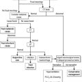
The aim is to treat the acute pain and then prevent chronic relapse with hypo-uricaemic drugs.
Acute attack
Colchicine
When NSAIDs are contraindicated, colchicine is used. Doses of colchicine of 0.5 mg 6- or 8-hourly orally have equivalent efficacy and a lower rate of gastrointestinal toxicity compared to higher doses.6 The higher doses such as colchicine 1.0 mg followed by 0.5 mg up to four times daily, with a maximum cumulative dose of 8 mg for an acute attack, are no longer recommended. All colchicine doses should be less with renal impairment, and may be restricted by the onset of nausea, vomiting and diarrhoea. Avoid prolonged colchicine use in patients with renal impairment as this may lead to a peripheral myoneuropathy.
Corticosteroids
Give patients with gout refractory to the above treatment or in whom both medications are contraindicated corticosteroids, such as prednisolone from 25 to 50 mg daily for 3 days then weaned over the course of 1 to 2 weeks.7,8 An alternative approach is to give intra-articular corticosteroid for monoarticular gout provided sepsis has been excluded. Educate all patients to correct lifestyle factors where appropriate.
Recurrent attacks
Urate lowering therapy
A second attack of gout usually requires urate lowering therapy, although this is not usually commenced in the emergency setting, as treatment must be delayed until the acute flare up has settled. Allopurinol, a xanthine oxidase inhibitor, prevents the production of uric acid from xanthine. It is introduced at a low dose once the acute attack has settled and gradually titrated up to a maximum of 300 mg daily.9 Typically, the patient will remain on a low-dose NSAID or colchicine (and/or prednisolone) as prophylaxis against precipitating further acute attacks. An alternative uricosuric agent to allopurinol is probenecid which should be avoided in renal impairment.
Management
Give oral analgesics and NSAIDs similar to acute gout for polyarticular pseudogout, as performing multiple joint injections is also impractical and painful. Take great care using NSAIDs in the elderly, and choose the smallest doses to avoid renal impairment and precipitating heart failure.
Summary of the approach to the management of acute monoarthritis
The British Society for Rheumatology in conjunction with other medical associations published guidelines in 2006 regarding an approach to the hot swollen joint.10 These guidelines are summarized below:
1 Gupta MN, Sturrock RD, Field M, et al. Prospective comparative study of patients with culture proven and high suspicion of adult onset septic arthritis. Annals of Rheumatic Diseases. 2003;62:327-331.
2 Jeng GW, Wang CR, Liu ST, et al. Measurement of synovial tumour necrosis factor-alpha in diagnosing emergency patients with bacterial arthritis. American Journal of Emergency Medicine. 1997;15:626-629.
3 Margaretten ME, Kohlwes J, Moore D, et al. Does this adult patient have septic arthritis? Journal of American Medical Association. 2007;297:1478-1488.
4 Sharma M, Leirisalo-Repo M. Arthritis patient as an emergency case at a university hospital. Scandinavian Journal of Rheumatology. 1997;26:30-36.
5 Zhang W, Doherty M, Pascual E, et al. EULAR evidence based recommendations for gout. Part 1: Diagnosis. Annals of Rheumatic Diseases. 2006;65:1301-1311.
6 Morris I, Varughese G, Mattingly P, et al. Colchicine in acute gout. British Medical Journal. 2003;327:1275-1276.
7 Cronstein BN, Terkeltaub R. The inflammatory process of gout and its treatment. Arthritis Respiratory Therapy. 2006;8(suppl 1):S3.
8 Rheumatology Guidelines, Version 1. Melbourne: Therapeutic Guidelines Ltd, 2006.
9 Zhang W, Doherty M, Bardin T, et al. EULAR evidence based recommendations for gout. Part 1: Management. Annals of Rheumatic Diseases. 2006;65:1312-1324.
10 Coakley G, Mathews C, Field M, et al. BSR and BHPR, BOA, RCGP and BSAC guidelines for management of the hot swollen joint in adults. Rheumatology. 2006;45:1039-1041.
Antibiotic Guidelines, Version 13. Melbourne: Therapeutic Guidelines Ltd, 2006.
Hochberg MC, et al. Rheumatology, 3rd edn. London: Elsevier, 2003.
Terkeltaub RA. Gout. New England Journal of Medicine. 2003;349:1647-1655.
UpToDate online. http://www.utdol.com/utd/content/search.do. accessed August 2008
14.3 Polyarticular rheumatism
ACUTE POLYARTHRITIS
Polyarthritis syndromes may be difficult to diagnose accurately due to the wide range of differential diagnoses, as shown in Table 14.3.1. Important measures are to rule out infection, quantify underlying inflammation and document extra-articular involvement.
Table 14.3.1 Differential diagnosis of polyarthritis syndromes1
| Inflammatory |
|---|
| Non-inflammatory |
|---|
Diagnosis and clinical features
History2,3
Take a focused history to include the following.
Mode of onset
Examination4
Perform a detailed physical examination and document:
Investigations1,2
Laboratory studies
Send serum antibody or antigen tests as indicated by the history including for infectious exposure such as hepatitis B serology, streptococcal antigen test and an auto-antibody panel including antinuclear antibody (ANA), rheumatoid factor and antibodies against cyclic citrullinated peptide (anti-CCP). Antibody tests in particular should be interpreted with caution and interpreted in the context of each individual patient, due to their varying sensitivities and specificities.5
RHEUMATOID ARTHRITIS
Rheumatoid arthritis (RA) is a chronic systemic inflammatory disorder of unknown aetiology characterized by symmetric synovitis, erosive polyarthritis and numerous extra-articular manifestations. It occurs in up to 2% of the general population and is two to three times more common in women.6 Its onset is often indolent, and may lack the characteristic symmetry of joint involvement. Uncommonly it presents as an acute monoarthritis.
Diagnosis
The diagnosis in adults requires four or more of the American College of Rheumatology criteria:7
Constitutional features such as malaise and fatigue are common.
Clinical features
Characteristic presentations in RA include the following.8–10
Extra-articular manifestations
The extra-articular manifestations of RA are protean, and may involve any organ system due to local inflammation causing functional or neurological deficits, rheumatoid vasculitis or distant inflammation. Patients may also present with the side effects of the treatment, including sepsis related to immunosuppression. Sepsis with encapsulated organisms is of particular concern in patients with Felty’s syndrome of RA, neutropenia and splenomegaly.11
Investigations
Laboratory studies
Send blood for FBC and ELFTs and non-specific markers of inflammation such as ESR, and CRP, with assays for serum rheumatoid factor and anti-CCP.12 Anti-CCP is less sensitive but more specific than rheumatoid factor for RA, and is more frequently positive early in the disease process. It is also thought to identify individuals at higher risk of erosive disease.13 Send blood cultures as well as mid-stream urine for suspected sepsis.
Imaging
Initial plain imaging of affected joints at first presentation does not usually demonstrate erosive changes, but is useful in patients with longstanding disease. However, in any patient with any cervical or neurological features, always request plain X-rays of the cervical spine to look for an atlanto-dens interval of greater than 2.5 mm, which is diagnostic of instability.14 Include a chest X-ray if there is a fever and/or any respiratory features. Request an ultrasound examination to differentiate deep vein thrombosis from a Baker’s cyst.
Emergency management
Medication falls broadly under the categories of NSAIDs and disease-modifying anti-rheumatic drug (DMARD) therapy. Readers are referred to Chapter 14.1 for a brief overview of these medications and common adverse effects, as well as the references at the end of this chapter.15–18
SERONEGATIVE ARTHROPATHIES
The seronegative arthropathies are characterized by inflammation of the axial spine with sacroiliitis and spondylitis in particular, enthesopathy which is inflammation at the attachments of tendons and ligaments to bones, dactylitis, asymmetric polyarthritis, eye inflammation often of the lower limb and varied mucocutaneous features.21 They are labelled ‘seronegative’ as serum rheumatoid factor is typically absent.
Epidemiology
However, the exact role of HLA-B27 in the pathogenesis of the spondyloarthropathies has not been clearly defined. The proportion of HLA-B27 positive individuals who develop symptomatic arthropathy varies widely from 16% in patients with AS to 70% of patients with spondylitis in the setting of IBD.22 HLA-B27 positive individuals may be less efficient at the intracellular removal of certain inciting bacteria, although this is controversial.23
PSORIATIC ARTHROPATHY
Psoriatic arthropathy is a heterogeneous disease with an identity distinct from other inflammatory arthritides. It occurs in 10% of patients with psoriasis, but may affect up to 40% of hospitalized psoriasis patients with widespread skin involvement.25 It occurs between the ages of 30 years and 60 years, with an equal prevalence in males and females. It is thought to be inherited in a polygenic pattern that is significantly influenced by environmental factors including trauma and infectious agents. Multiple studies have confirmed the important role of class I HLA, particularly B13, B16 and B27 and certain C-subclasses.26,27 The arthropathy pattern may be pauci-articular, but more than five peripheral joints are usually involved.
Diagnosis and clinical features
The diagnosis of psoriatic arthropathy is essentially clinical, requiring the demonstration of coexisting synovitis and psoriasis. A set of simple clinical diagnostic criteria (abbreviated to the CASPAR criteria) were recently proposed by a large international study group.28
The CASPAR diagnostic criteria for psoriatic arthropathy
Established inflammatory joint disease, and at least three points from the following features:
Five clinical subtypes are recognized, including asymmetric oligoarthritis, symmetric small joint polyarthritis, predominant distal interphalangeal joint involvement, psoriatic spondyloarthropathy and arthritis mutilans.29 Major extra-articular organ manifestations such as aortic insufficiency and pulmonary fibrosis occur rarely. However, up to 30% of patients have mild inflammation at the eye, most commonly conjunctivitis.
Asymmetric oligoarthritis
This occurs in 30–50% of patients.30 It presents as an oligoarthritis involving a single large joint, in association with a ‘sausage-shaped’ or dactylitic digit or toe. Dactylitis occurs due to a combination of arthritis and tenosynovitis. Distal interphalangeal joint involvement is typical, and is almost invariably associated with psoriatic nail changes of pitting, ridging and onycholysis. Enthesopathy occurs most frequently with this form of the disease, and commonly manifests as plantar fasciitis or epicondylitis at the elbow.
Symmetric small joint polyarthritis
This occurs in 30% of patients, in a pattern strongly resembling RA, but with more frequent distal interphalangeal joint involvement.30
Psoriatic spondyloarthropathy
This occurs in 5% of patients.30 It is often asymptomatic, but may present with inflammatory low back pain due to sacroiliitis in up to 30% of cases.
Arthritis mutilans
‘Arthritis mutilans’ is a rare (<5% of patients), but well-characterized feature of psoriatic arthritis, with severely deforming arthritis including telescoping of the fingers or toes from osteolysis of the metacarpal or metatarsal bones and phalanges.30
Dermatological features
Dermatological features include typical erythematous, scaling plaques on the extensor surfaces of the elbows and knees, scalp and ears, and nail changes. The nail changes include pitting with usually greater than 20 pits, ridging with transverse depressions, and onycholysis with separation of the nail from the underlying nail bed.25 Nodules and vasculitic features such as digital ulcers do not occur.
Investigations
ESR and CRP are raised, but the rheumatoid factor and autoantibody screen are negative. Plain imaging studies of affected joints may reveal typical radiographic features, including soft tissue swelling, bone proliferation at the base of digital phalanges coupled with resorption of the distal tufts (the ‘pencil-in-cup’ deformity) and fluffy periostitis.31 Chest radiographs are useful as a baseline when clinical examination suggests cardiac or pulmonary involvement.
Emergency and ongoing management
NSAIDs are useful for acute symptomatic relief, and intra- or peri-articular corticosteroids may be used for short-term relief of painful arthritis or enthesitis. Long-term therapy with disease modifying agents, such as sulphasalazine or methotrexate, is instituted at specialist review.32 Oral corticosteroids are usually avoided, as their cessation often exacerbates the psoriasis. Therapy with tumour necrosis factor α antagonists32 such as infliximab or etanercept has recently been approved to rheumatologists under strict access criteria for severe disease resistant to other DMARD therapy.
Emergency management of skin disease includes topical treatments such as emollients and keratolytic agents.33 Phototherapy and photo-chemotherapy may be instituted on early dermatological consultation. Admit patients if their symptoms are severe enough to preclude oral therapy or safe discharge pending outpatient specialist follow-up.
Prognosis
Psoriatic arthropathy generally runs a more benign course than RA, but patients nonetheless suffer from considerable morbidity. Adverse prognostic factors include onset before 20 years of age, erosive disease and extensive skin involvement.30
REACTIVE ARTHRITIS
Reactive arthritis is aseptic peripheral arthritis following certain infections, which include bacterial infections of the urogenital tract usually by Chlamydia trachomatis, or of the gastrointestinal tract with organisms such as Shigella, Salmonella and Campylobacter. It may also follow viral infections such as HIV, although in the case of HIV, coinfection with sexually transmitted organisms rather than the virus itself is thought to cause the symptoms.34 The seroconversion illness of HIV with its own constellation of articular symptoms is considered to be a separate entity.
Epidemiology
The prevalence of reactive arthritis has been difficult to define owing to diagnostic uncertainty particularly in the setting of asymptomatic sexually transmitted infection. The male preponderance is up to 9:1 following sexually transmitted infection, but males and females are equally affected following gastrointestinal tract infection.35 The peak incidence is around the age of 35 years, and up to 75% of patients are HLA-B27 positive.35 An important exception is with the reactive peripheral arthritis that occurs in 20% of patients with idiopathic IBD, a condition that may mimic gastrointestinal tract infection, but where patients are usually HLA-B27 negative.
Diagnosis and clinical features
The diagnosis of reactive arthritis is clinical. It typically manifests within a month of gastrointestinal or genitourinary infection, although the latter is frequently asymptomatic.36 Musculoskeletal manifestations include myalgias and asymmetric polyarthritis affecting the knees, ankles and small joints of the feet in particular, although peripheral upper limb involvement is seen. Affected joints demonstrate marked inflammatory features with erythema, swelling, warmth and exquisite pain on active or passive movement. Fever and malaise are common.
Arthropathy and extra-articular manifestations
Symptomatic spondylitis and sacroiliitis cause low back and buttock pain and occur frequently. Dactylitis and enthesopathy are characteristic features of this disease with heel pain from plantar fasciitis or Achilles tendonitis.36
Extra-articular features associated with reactive arthritis include keratoderma blennorrhagicum; the scattered, thickened, hyperkeratotic skin lesions with pustules and crusts seen in Reiter syndrome, and circinate balanitis. An inflammatory aortitis occurs in 1% of patients and may result in aortic valvular incompetence, and/or heart block. Keratoderma blennorhagicum on the soles or palms may coalesce to form plaques virtually indistinguishable from those of psoriasis.37 Circinate balanitis causes shallow meatal ulcers that are moist in uncircumcised men or hyperkeratotic and plaque-like in circumcised men.37
The peripheral arthritis associated with IBD is migratory and occurs in a similar distribution. Common features include large joint effusions, particularly involving the knee, and sacroiliitis or spondylitis.38 Unlike peripheral arthritis following genitourinary infection, the spondylitis of IBD-associated arthropathy does not tend to settle with treatment of the bowel inflammation. Cutaneous features associated with this form of arthropathy occur mainly on the lower limbs, and include erythema nodosum and pyoderma gangrenosum.38
Investigations
Blood testing
An active inflammatory response is seen in the acute phase with a neutrophil leukocytosis and thrombocytosis, and raised ESR and CRP. The presence of a mild normochromic, normocytic anaemia suggests chronic disease. Send blood for HLA-B27. Document the preceding genitourinary or gastrointestinal organism by stool culture or cervical/urethral swabs.39 Rheumatoid factor and ANA are negative.
Joint aspiration
Joint aspiration may be necessary to exclude intra-articular sepsis (see Ch. 14.2). The synovial fluid may be turbid, viscous and with a neutrophil leukocytosis up to 50 000/mm3, but Gram stain and bacterial culture are negative, and unlike true septic arthritis, the synovial glucose level is not significantly reduced compared to serum levels.39 Macrophages with intracytoplasmic vacuoles containing ingested neutrophils are occasionally seen.
Emergency and ongoing management
Give antibiotics such as doxycycline 100 mg orally bd for 7 days or azithromycin 1 g orally once for documented urethritis or cervicitis, and remember partner contact tracing and treatment. An infectious diseases opinion is useful in these cases.39
Admit patients with suspected septic arthritis until it is excluded, or if they are unable to tolerate simple oral therapies. Request a cardiology opinion for major cardiac involvement with valvular disease or a conduction abnormality, and a gastroenterology opinion when IBD is suspected, although the role of treatment and the effect on the arthropathy is unclear. Multidisciplinary physical therapy is essential on an outpatient basis.
Prognosis
Signs and symptoms usually remit within 6 months. However, up to 50% of patients suffer from recurrent arthritis, and up to 30% develop chronic arthropathy.40 Post-dysenteric cases have a better prognosis than post-chlamydial cases. Poor prognostic signs include early onset under the age of 16 years, hip involvement and the presence of dactilytis.
POLYARTICULAR CRYSTAL ARTHROPATHY
Crystal-induced arthropathies result from the deposition of crystal in joint spaces, such as in gout or pseudogout. Both diseases cause debilitating joint inflammation resulting from the lysis of neutrophil polymorphs that have ingested monosodium urate in the case of gout or calcium pyrophosphate crystals in pseudogout. Although usually monoarticular, polyarticular involvement can occur in up to 5% of cases. See Chapter 14.2 for a detailed discussion of these diseases.
Viral arthritis
Alphaviruses
Alphaviruses are a mosquito-borne genus of the Togaviridae family. They are responsible for epidemics of febrile polyarthritis, including Ross River, Barmah Forest and Sindbis viruses in Australia, West Nile virus that has recently been documented in the USA, Chikungunya virus in East Africa, South and South-East Asia, O’nyong-nyong virus in East Africa and Mayaro virus in South America.41
Ross River virus
RRV is endemic to Australia, New Zealand and South Pacific islands, and is the most common arboviral disease in Australia. RRV is transmitted by the Ochlerotatus (formerly Aedes) vigilex mosquito via a marsupial reservoir.42 Epidemics of acute febrile polyarthritis are most common between January and May, but can occur after periods of heavy rains.
Diagnosis and clinical features of Ross River virus
A detailed travel history is essential. There is usually low grade fever and other constitutional symptoms. A rash varying in distribution, character and duration occurs up to 2 weeks before, during or after the other symptoms. Polyarticular symptoms are present in most patients with a symmetric arthritis or arthralgia primarily affecting the wrist, knee, ankle and small joints of the extremities. Cervical lymphadenopathy occurs frequently, and paraesthesiae and tenderness of the palms and soles in a small percentage of cases.43
Investigations
Serology for Ross River virus
Serology testing distinguishes RRV from other causes of febrile polyarthritis such as Barmah Forest virus. A significant rise in IgM antibody titre to RRV indicates acute infection, or the virus itself may be isolated from the serum of acutely unwell patients. Radiographs are unremarkable and uneconomic as the disease is largely self-limiting.44
Emergency and ongoing management
Patients with RRV require symptomatic treatment with simple analgesics or NSAIDs. Occasionally, a brief course of low-dose prednisolone may be used. RRV is a notifiable disease.42 Conventional personal preventative measures such as protective clothing, effective mosquito repellent and avoidance of mosquito-prone areas should be recommended, as no vaccine currently exists. Refer to a rheumatologist if symptoms are severe or refractory to simple treatment measures.
Parvovirus B19
Human parvovirus B19 infection is caused by a small, single-stranded DNA virus that has a predilection for erythroid precursor cells and is transmitted by respiratory secretions. It causes the self-limiting illness Erythema infectiosum or ‘slapped cheek disease’ or ‘fifth disease’ in children. In adults, however, parvovirus B19 manifests with severe flu-like symptoms, and as many as 75% develop joint symptoms. It may be responsible for up to 12% of adult patients presenting with acute polyarthritis, most notably in those who have frequent exposure to children.45
Diagnosis and clinical features
The characteristic rash is usually absent in adults. An acute polyarthritis improves over 2 weeks, with symmetric involvement of peripheral small joints, including the hands (proximal interphalangeal and metacarpophalangeal joints in particular), wrists, knees and ankle joints. Morning stiffness is a prominent feature. These features are similar to those seen in patients with RA, and in fact up to 50% of affected patients meet the ACR diagnostic criteria for RA.46 Uncommon but important extra-articular features of parvovirus B19 infection are detailed below.47
Investigations
Always send an FBC, particularly given the potential for an aplastic crisis and bone marrow suppression. Non-specific markers of inflammation are likely to be elevated. Specific serological diagnosis is made by demonstrating high IgM antibody titres specific to the virus, and by isolation of the viral DNA by polymerase chain reaction (PCR). IgG antibodies to parvovirus B19 indicate past infection and are common in the adult population.48
Transient, moderate elevations of rheumatoid factor, anti-DNA, antilymphocyte or anticardiolipin antibodies sometimes occur.48 Radiographs of the affected joints are normal.
Emergency and ongoing management
Rest and NSAIDs are the mainstay of emergency treatment, except in pregnant women, as NSAIDs are contraindicated in the third trimester.49 A short course of prednisolone may be required. Significant extra-articular manifestations may require admission and consultation with the appropriate specialist. Blood transfusion or intravenous immunoglobulin infusions may be necessary.
Prognosis
Joint symptoms are self-limited in the majority of adult patients, but up to 10% may have prolonged relapsing and remitting symptoms lasting up to 9 years.50
Hepatitis B and Hepatitis C Viruses
The common hepatitis viruses A, B and C all cause viral polyarthritis. Hepatitis B virus (HBV) is responsible for 20–25%, and hepatitis A (HAV) up to 14% of causes in patients with viral polyarthritis.51 The polyarthritis of HAV tends to occur during the infectious phase, and is self-limiting. The polyarthritis of HBV occurs in early infection during a period of significant viraemia, and is thought to be due to immune complexes.
Diagnosis and clinical features
HBV polyarthritis is acute and severe, and manifests in a symmetric, migratory or additive fashion most commonly involving the hand and knee joints.52 Other large axial joints may be involved, and significant early morning stiffness is often present. The arthritis may precede the development of jaundice, and persist for several weeks after jaundice has developed.
Hepatitis C virus (HCV) polyarthritis is rapidly progressive and symmetrical, involving the hands, wrists, shoulders, knees and hips.53 Carpal tunnel syndrome and tenosynovitis may occur. It is unusual for polyarthritis to be the first manifestation of the underlying disease in either HBV or HCV. Nonetheless, ask about exposure risk factors for these viruses such as intravenous drug abuse, unprotected sexual intercourse, past blood transfusions, tattoos, as well as about previous jaundice. Both hepatitis B and hepatitis C disease are associated with a number of important extra-articular, extra-hepatic manifestations.
Emergency and ongoing management
Commence symptomatic treatment of the polyarthritis with NSAIDs in the emergency department. Refer refractory HBV- or HCV-associated polyarthritis to a rheumatology specialist and/or combined hepatology clinic. Disease modifying agents such as prednisolone and sulphasalazine may be used cautiously with careful monitoring of the liver function tests and for increasing viraemia.56
RHEUMATIC FEVER
Acute rheumatic fever (ARF) refers to a constellation of non-infectious symptoms occurring after a pharyngeal infection with group A streptococci (GAS). Anecdotal evidence suggests that it may also occur in high-risk populations following skin infections with GAS.57
Epidemiology
ARF is characterized by inflammation of connective tissue including the joints, subcutaneous tissue, heart and blood vessels. Its prevalence has declined over time in developed countries, but it remains a major public health problem in developing countries, and in the more socially isolated parts of Australasia. The highest documented rates in the world occur in the Aboriginal Australian population and Torres Strait Islander populations of New Zealand and the Pacific Islands.58
ARF is primarily a disease of children aged 5–14 years. The annual incidence may reach 350 per 100 000 in Aboriginal children.59 However, the polyarthritis of ARF is most commonly seen in adolescents and young adults.
Diagnosis and clinical features
The diagnosis of ARF worldwide is made on the 1944 Jones or more recently World Health Organization major and minor criteria. However, these criteria appear too restrictive for diagnosing ARF in Australian indigenous populations. Therefore, new criteria for use in high- and low-risk populations in Australia have been proposed (see Table 14.3.2).60
Table 14.3.2 2005 Australian guidelines for the diagnosis of acute rheumatic fever
| High-risk groups | All other groups | |
|---|---|---|
| Initial episode of ARF | Two major or one major and two minor manifestations plus evidence of a preceding GAS infection | Two major or one major and two minor manifestations plus evidence of a preceding GAS infection |
| Recurrent attack of ARF in a patient with known past ARF or RHD | Two major or one major and two minor or three minor manifestations plus evidence of a preceding GAS infection | Two major or one major and two minor or three minor manifestations plus evidence of a preceding GAS infection |
| Major manifestations |
ARF, acute rheumatic fever; GAS, group A streptococci; RHD, rheumatic heart disease.
The polyarthritis of ARF is usually the earliest symptom of the disease, and is classically described as migratory affecting several joints in quick succession for a short time, commencing with the large joints of the lower limb then the large joints of the upper limb.61 Affected joints are painful but objective signs of inflammation such as erythema and swelling are not prominent.
Fever and constitutional symptoms are common. Other important extra-articular major criteria (with polyarthritis) of the disease include the following.61
Major criteria (with polyarthritis) of acute rheumatic fever
Investigations
Measure antistreptolysin O and antideoxyribunuclease B (anti-DNase B) titres.61 As these titres can take 6 weeks after infection to peak, interpretation in the acute phase should be cautious, and serial tests should be performed. Note that anti-streptococcal antibody titres are useful in low-risk populations, but are difficult to interpret in high-risk populations due to pre-existing high background titres.62 Send a throat swab, although this is positive in less than 10% of high-risk populations. Other important tests include:
Emergency and ongoing management
Emergency management of ARF depends on making the diagnosis, and treating the manifestations. Patients are severely symptomatic, and often require admission for initial observation and management. Request rheumatology and infectious diseases opinions and a neurology opinion if chorea is troublesome. The presence of heart block or, more importantly, frank cardiac failure or acute valvular regurgitation mandate cardiac admission.
The polyarthritis of rheumatic fever is exquisitely responsive to NSAID therapy, particularly aspirin, so much so that failure of NSAID therapy to rapidly relieve symptoms should prompt consideration of an alternative diagnosis.63 Give aspirin at doses of 80–100 mg/kg/day in 4–5 divided doses in adults, usually for 1 to 2 weeks.64
Prognosis
Recurrence of ARF commonly occurs within 2 years of the initial attack, despite prophylactic therapy. Most affected connective tissues do not sustain long-lasting damage, with the exception of the heart, which is prone to additive subclinical damage resulting in rheumatic heart disease.65
1 Klinkhoff A. Rheumatology: 5. Diagnosis and management of inflammatory polyarthritis. Canadian Medical Association Journal. 2000;162:1833-1838.
2 Pinals RS. Polyarthritis and fever. New England Journal of Medicine. 1994;330:769.
3 Richie A, Francis M. Diagnostic approach to polyarticular joint pain. American Family Physcian. 2003;68(6):1075-1088.
4 Talley NJ, O’Connor S. The rheumatological system. In: Talley NJ, O’Connor S, eds. Clinical examination: a systematic guide to physical diagnosis. 5th edn. Sydney: Churchill Livingstone.
5 Woolf SH, Kamerow DB. Testing for uncommon conditions. The heroic search for positive test results. Archives of Internal Medicine. 1990;150:2451. 2058
6 Rindfleisch JA, Muller D. Diagnosis and management of rheumatoid arthritis. American Family Physician. 2005;72(6):1037-1047.
7 Arnett FC, Edworthy SM, Bloch DA, et al. The American Rheumatism Association 1987 revised criteria for the classification of rheumatoid arthritis. Arthritis and Rheumatism. 1988;31:315-324.
8 Rindfleisch JA, Muller D. Diagnosis and management of rheumatoid arthritis. American Family Physician. 2005;72(6):1037-1047.
9 Scott DL. Rheumatoid arthritis: acute presentations and urgent complications. British Journal of Hospital Medicine. 2006;67(5):235-239.
10 Tutuncu Z, Kavanaugh A. Rheumatic disease in the elderly: rheumatoid arthritis. Rheumatic Diseases Clinics of North America. 2007;33(1):57-70.
11 Balint GP, Balint PV. Felty’s syndrome. Best Practices and Research Clinics Rheumatology. 2004;18(5):631-645.
12 Westwood OM, Nelson PN, Hay FC. Rheumatoid factors: what’s new? Rheumatology. 2006;45:379-385.
13 American College of Rheumatology. The use of anti-cyclic citrullinated peptide (anti-CCP) antibodies in RA, 2003. http://www.rheumatology.org/publications/hotline/1003anticcp.asp. (accessed August 2008)
14 Macarthur A, Kleiman S. Rheumatoid cervical joint disease – a challenge to the anaesthetist. Canadian Journal of Anaesthesia. 1993;40(2):154-159.
15 Emery P. Treatment of rheumatoid arthritis. British Medical Journal. 2006;332(7534):152-155.
16 O’Dell JR. Therapeutic strategies for rheumatoid arthritis. New England Journal of Medicine. 2004;350(25):2591-2602.
17 Strand V, Hochberg MC. The risk of cardiovascular thrombotic events with selective cyclooxygenase-2 inhibitors. Arthritis and Rheumatism. 2002;47:349-355.
18 Olsen NJ, Stein CM. New drugs for rheumatoid arthritis. New England Journal of Medicine. 2004;350:2167-2179.
19 Alarcon GS. Predictive factors in rheumatoid arthritis. American Journal of Medicine. 1997;103(6A):19S-24S.
20 Wagner U, Kaltenhauser S, Sauer H, et al. HLA markers and prediction of clinical course and outcome in rheumatoid arthritis. Arthritis and Rheumatism. 1997;40(2):341-351.
21 Young JL, Smith L, Matyszak MK. HLA-B27 Expression does not modulate intracellular Chlamydia trachomatis infection of cell lines. Infection and Immunity. 2001;69(11):6670-6675.
22 Dougados M, van der Linden S, Juhlin R, et al. The European Spondylarthropathy Study Group preliminary criteria for the classification of spondylarthropathy. Arthritis and Rheumatism. 1991;34(10):1218-1227.
25 Myers WA, Gottlieb AB, Mease P. Psoriasis and psoriatic arthritis: clinical features and disease mechanisms. Clinics in Dermatology. 2006;24(5):438-447.
26 Eastmond CJ. Psoriatic arthritis. Genetics and HLA antigens. Baillières Clinical Rheumatology. 1994;8(2):263-276.
27 Espinoza LR, van Solingen R, Cuellar ML, et al. Insights into the pathogenesis of psoriasis and psoriatic arthritis. American Journal of Medical Science. 1998;316(4):271-276.
28 Taylor W, Gladman D, Helliwell P. Classification criteria for psoriatic arthritis: development of new criteria from a large international study. Arthritis and Rheumatism. 2006;54(8):2665-2673.
29 Cuellar ML, Silveira LH, Espinoza LR. Recent developments in psoriatic arthritis. Current Opinion of Rheumatology. 1994;6(4):378-384.
30 Mease P, Goff BS. Diagnosis and treatment of psoriatic arthropathy. Journal of the American Academy of Dermatology. 2005;52(1):1-19.
31 Ory PA, Gladman DD, Mease PJ. Psoriatic arthritis and imaging. Annals of Rheumatic Diseases. 2005;2(64 suppl):ii55-ii57.
32 Mease P. Management of psoriatic arthritis: the therapeutic interface between rheumatology and dermatology. Current Rheumatology Reports. 2006;8(5):348-354.
33 Menter A, Griffiths CE. Current and future management of psoriasis. Lancet. 2007;370(9583):272-284.
34 Hamdulay SS, Glynne SJ, Keat A. When is arthritis reactive? Postgraduates in Medicine Journal. 2006;82(969):446-453.
35 Toivanen A, Toivanen P. Reactive arthritis. Best practice and research. Clinical Rheumatology. 2004;18(5):689-703.
36 Amor B. Reiter’s syndrome. Diagnosis and clinical features. Rheumatic Diseases Clinics of North America. 1998;24(4):677-695.
37 Angulo J, Espinoza LR. The spectrum of skin, mucosa and other extra-articular manifestations. Baillières Clinical Rheumatology. 1998;12(4):649-664.
38 Holden W, Orchard T, Wordsworth P. Enteropathic arthritis. Rheumatic Diseases Clinics of North America. 2003;29(3):513-530. viii
39 Petersel DL, Sigal LH. Reactive arthritis. Infectious Disease Clinics of North America. 2005;19(4):863-883.
40 Colmegna I, Espinoza LR. Recent advances in reactive arthritis. Current Rheumatological Reports. 2005;7(3):201-207.
41 Suhrbier A, La Linn M. Clinical and pathologic aspects of arthritis due to Ross River virus and other alphaviruses. Current Opinion in Rheumatology. 2004;16(4):374-379.
42 Ross River virus infection Factsheet. Australian Government Department of Health and Ageing, May 2004. http://www.health.gov.au/. (accessed August 2008)
43 Mylonas AD, Brown AM, Carthew TL, et al. Natural history of Ross River virus-induced epidemic polyarthritis. Medical Journal of Australia. 2002;177(7):356-360.
44 Cheong IR. Ross River virus: are we wasting money doing tests? Medical Journal of Australia. 2003;178(3):143.
45 Servey JT, Reamy BV, Hodge J. Clinical presentations of parvovirus B19 infection. American Family Physician. 2007;75(3):373-376.
46 Naides SJ, Scharosch LL, Foto F, et al. Rheumatologic manifestations of human parvovirus B19 infection in adults. Initial 2-year clinical experience. Arthritis and Rheumatism. 1990;31:1297-1309.
47 Calabrese LH, Naides SJ. Viral arthritis. Infectious Disease Clinics of North America. 2005;19(4):963-980. x
48 Moore TL. Parvovirus-associated arthritis. Current Opinion in Rheumatology. 2000;12(4):289-294.
49 Jacqz-Aigrain E, Koren G. Effects of drugs on the fetus. Seminars in Fetal and Neonatal Medicine. 2005;10(2):139-147. Epub 2005 Jan 25
50 Broliden K, Tolfvenstam T, Norbeck O. Clinical aspects of parvovirus B19 infection. Journal of Internal Medicine. 2006;260(4):285-304.
51 Franssila R, Hedman K. Infection and musculoskeletal conditions: viral causes of arthritis. Best Practices and Research Clinics of Rheumatology. 2006;20(6):1139-1157.
52 Chi ZC, Ma SZ. Rheumatologic manifestations of hepatic diseases. Hepatobiliary and Pancreat Diseases International. 2003;2(1):32-37.
53 Sanzone AM, Bégué RE, Hepatitis C. and arthritis: an update. Infectious Disease Clinics of North America. 2006;20(4):877-889. vii
54 Farrell GC, Teoh NC. Management of chronic hepatitis B virus infection: a new era of disease control. International Medicine Journal. 2006;36(2):100-113.
55 Hoofnagle JH. Course and outcome of hepatitis C. Hepatology. 2002;36(5 Suppl 1):S21-S29.
56 Ramos-Casals M, Trejo O, García-Carrasco M. Therapeutic management of extrahepatic manifestations in patients with chronic hepatitis C virus infection. Rheumatology (Oxford). 2003;42(7):818-828. Epub 2003 Apr 16
57 McDonald M, Currie B, Carapetis J. Acute rheumatic fever: a chink in the chain that links the heart to the throat? Lancet Infectious Diseases. 2004;4:240-245.
58 Carapetis JR, et al. The global burden of group A streptococcal diseases. Lancet Infectious Diseases. 2005;5:685-694.
59 Australian Institute of Health and Welfare. Rheumatic heart disease in Australia – all but forgotten except in Aboriginal and Torres Strait Islander peoples. Canberra: AIHW, 2004.
60 National Heart Foundation of Australia (RF/RHD guideline development working group) and the Cardiac Society of Australia and New Zealand.Diagnosis and management of acute rheumatic fever and rheumatic heart disease in Australia – an evidence based review. National Heart Foundation of Australia, 2006; 6-24. http://www.heartfoundation.org.au/Professional_Information/Clinical_Practice/ARF_RHD.html. accessed August 2008
61 Carapetis JR, McDonald M, Wilson NJ. Acute rheumatic fever. Lancet. 2005;366(9480):155-168.
62 Nimmo GR, Tinniswood RD, Nuttall N, et al. Group A streptococcal infection in an Aboriginal community. Medical Journal of Australia. 1992;156:537-540.
63 Cilliers AM. Rheumatic fever and its management. British Medical Journal. 2006;333(7579):1153-1156.
64 Thatai D, Turi DG. Current guidelines for the treatment of acute rheumatic fever. Drugs. 1999;57:545-555.
65 Figueroa FE, Fernández MS, Valdés P. Prospective comparison of clinical and echocardiographic diagnosis of rheumatic carditis: long term follow-up of patients with subclinical disease. Heart. 2001;85(4):407-410.
14.4 Musculoskeletal and soft tissue emergencies
Common causes of soft tissue injuries


General evaluation of a soft tissue injury
Puncture injuries
Clinical evaluation
This type of injury is caused by stepping on a nail or pin, penetration by a sewing machine needle, or industrial equipment such as a nail gun, high pressure hose or gun. Needlestick and sharps incidents are covered in Chapter 9.10.
Acute mechanical overload injuries
These include fractures, ligament sprains, muscle strains or tears and tendon ruptures. Many are covered in Section 4.
Classification of ligament sprains/muscle strains
| Grade I | Small number of fibres injured, with pain on stressing, but no laxity or loss of strength. |
| Grade II | Significant number of fibres injured with laxity and or weakness and pain on stressing. |
| Grade III | Complete tear with gross laxity and no strength. |
Degloving injuries
Evaluation
Closed degloving injuries are harder to diagnose and may only lead to the skin feeling slightly less tethered than prior to injury, a failure to blanch with pressure, poor capillary return and most importantly altered cutaneous sensation. This is most easily and accurately assessed by 2-point discrimination. If 2-point discrimination is normal, a significant degloving injury has not occurred. Pain may or may not be prominent, and/or may relate to an underlying bony injury.
Chronic overload (overuse) injuries
Classification
Classification of chronic overuse syndromes
| Grade I | Pain after activity |
| Grade II | Pain early on and after activity; activity not limited |
| Grade III | Pain throughout activity, which is limited |
| Grade IV | Pain at rest. |
Calcaneus
Diagnosis and management
Stress fracture of the calcaneus causes the insidious onset heel pain, which is worse with weight bearing and is tender on lateral compression of the heel. It is most common in distance runners or in the military related to marching. X-ray is occasionally abnormal, but otherwise the diagnosis is made on a bone scan, CT scan or MRI. Arrange for a non-weight bearing POP cast for 6 weeks and referral to an orthopaedic specialist.
Non-articular rheumatism
Frozen shoulder (adhesive capsulitis)
Diagnosis
Frozen shoulder (adhesive capsulitis) has a natural history over 1–5 years, with an average duration of 2.5 years. It begins with an acutely painful period of 3–9 months, with progressively decreasing range of motion at the glenohumeral joint over 4–12 months that starts soon after the pain. The decreased range of motion of about 15–45 degrees of movement usually entirely resolves but takes from 1 to 4 years to do so. The pain tends to be worse at night or when lying flat.
Management
The only treatments with evidence of efficacy are high-dose intra-articular steroid injection that reduces early pain but without effect on the range of motion, and physical disruption of the joint capsule, for instance by arthroscopic capsule release. There is no evidence that NSAIDs or physiotherapy alone have any effect on outcome, and NSAIDs in particular have an increased risk of causing renal impairment, heart failure and gastrointestinal bleeding in the elderly (see Ch. 22.1).
Olecranon bursitis
Diagnosis and management
Refer the patient for drainage of the bursa under anaesthesia if significant bacterial infection is confirmed, or if a septic arthritis itself is suspected by markedly reduced movement at the elbow (see Ch. 14.2). Otherwise, give an antistaphylococcal antibiotic such as di- or flucloxacillin 500 mg orally qid for 7 to 10 days, and/or a non-steroidal anti-inflammatory analgesic, and refer back to the GP.
Prepatellar bursitis (Housemaid’s knee)
De Quervain’s stenosing tenosynovitis
Diagnosis and management
This causes tenderness over the radial styloid, a palpable nodule from thickening of the fibrous sheaths of the abductor pollicis longus and extensor pollicis brevis tendons, and pain on moving the thumb. Treat by resting the thumb in a splint and by using an anti-inflammatory analgesic.
Plantar fasciitis
Diagnosis and management
Plantar fasciitis presents as a painful midfoot, especially in the sole or arch, that is worse on first weight bearing and improves after 10–15 min of walking, recurring again during load bearing for an extended period. It is one of the most common causes of recurrent foot pain, and may be one manifestation of the spondyloarthropathy seen in Reiter syndrome, ankylosing spondylitis and psoriatic arthritis (see Ch. 14.3). On examination, there is tenderness of the plantar fascia especially at the calcaneal attachment.
Back pain
This is a common problem that may be considered under four major groups that include back pain following direct major trauma, or minor indirect mechanical trauma; and severe or atypical non-traumatic back pain, or mild to moderate non-traumatic back pain. Direct major thoracic and lumbosacral spine trauma is covered in Chapter 3.3.
Indirect mechanical back trauma
Clinical features
Examination
Assess for a reduced straight-leg raise (SLR), but if a patient is able to sit up in bed with the legs out straight, this is equivalent to an SLR of 90 ° on both sides. Examine for signs of nerve-root irritation or compression from an acute lumbar disc prolapse. See Chapter 3.3 for a description of the myotomes, dermatomes and nerve roots in the leg.
Look for motor loss occurring in the following myotomes:
Assess for any reflex loss, with the knee jerk (L3, L4) and the ankle jerk (L5, S1).
Check for sensory loss occurring in the following dermatomes:
Non-traumatic severe or atypical back pain
Management
Refer the patient to the appropriate specialist team according to the suspected aetiology. Never send home a patient with abnormal vital signs or an abnormal physical examination, or one who has failed an analgesia regime. Always consider the possibility of a serious cause and avoid diagnosing ‘musculoskeletal pain? cause.’ See Chapters 14.1, 14.2 and 14.3 for the management of associated rheumatological conditions.
Non-traumatic mild-to-moderate back pain
Diagnosis and management
Bolin D, Kemper A. Current Concepts in the Evaluation and Management of Stress Fractures. Current Sports Medicine Reports. 2005;4(6):295-300.
Booth C. High pressure paint gun injuries. British Medical Journal. 1977;2:1333-1335.
Brown AFT, Cadogan MD. Emergency medicine. Emergency and acute medicine: diagnosis and management, 5th edn. Hodder Arnold, London, 2006.
Brukner P, Khan K. Clinical Review of Sports Medicine, 2nd edn. Sydney: McGraw-Hill, 2001.
Mehallo C, Drezner J, Bytomski J. Practical Management. NSAID use in athletic injuries. Clinical Journal of Sport Medicine. 2006;16(2):170-174.
NHMRC Acute Pain Management: Scientific Evidence. ANZCA and Faculty of Pain Medicine, 2nd ed. 2005. www.nhmrc.gov.au/publications/synopses/cp104syn.htm/. Available at Accessed Nov 2008
NHMRC Evidence-Based Management of Acute Musculoskeletal Pain. A Guide for Clinicians. Australian Acute Musculoskeletal Pain Guidelines Group, 2004. Available at www.nhmrc.gov.au/publications/synopses/cp94syn.htm/ Accessed Nov 2008
Tatso J, Elias D. Adhesive Capsulitis. Sports Medicine and Arthroscopy Review. 2007;15(4):216-221.
UpToDate online version 15.2. http://www.utdol.com/utd/content/search.do. accessed August 2008