History: A 62-year-old woman, asymptomatic, is undergoing a screening examination.
1. Which of the following should be included in the differential diagnosis? (Choose all that apply.)
2. What is the treatment for the lesion demonstrated in the provided images?
D. No specific treatment is necessary
3. Which is the most common histological subtype of colonic adenomatous polyp?
4. What is the typical clinical presentation of a patient with a colonic polyp?
B. Bright rectal bleeding (hematochezia)
ANSWERS
CASE 14
Adenoma of the Colon
1. A, C, D, and E
2. C
3. B
4. E
References
O’Brien MJ, Winawer SJ, Zauber AG, et al: The National Polyp Study: patient and polyp characteristics associated with high-grade dysplasia in colorectal adenomas. Gastroenterology. 1990;98:371–379.
Cross-Reference
Gastrointestinal Imaging: THE REQUISITES, 3rd ed, p 268-271.
Comment
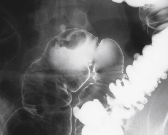
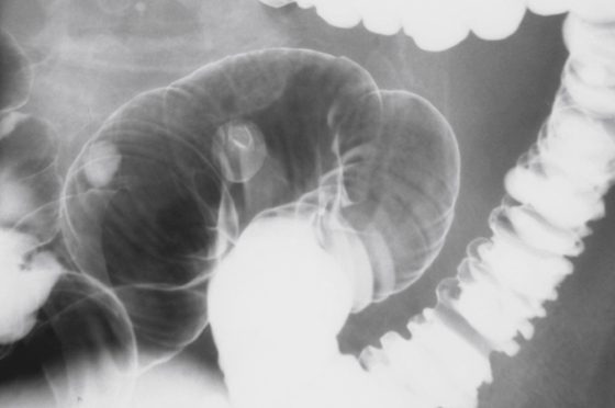
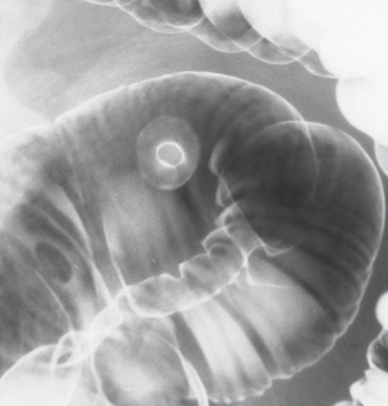
Colonic adenomas are found in the colons of 15% to 30% of patients older than 60 years, with the prevalence very dependent on age, heredity, and risk factors. Polyps come in three histologically identifiable forms. The first, and most common (80%), is the tubular adenoma. These polyps are usually small and carry very little risk for malignant degeneration. They can be on long stalks (see figures). However, the fact that the stalk permits the polyp to move with the patient can occasionally make detection on both air contrast barium enema (ACBE) and CT quite difficult.
The other two types are tubovillous adenomas and villous adenomas. Villous adenomas have the greatest risk for developing into a malignancy. Some researchers and clinicians consider a colonic villous adenoma a low-grade malignancy regardless of what the histologic picture shows. Polyps less than 0.5 mm (diminutive polyps) have little or no malignant potential. Polyps 1 to 2 cm in size carry about a 10% risk. Polyps greater than 2 cm are generally thought to have a 30% to 40% risk of malignancy.
The anatomic distribution of colonic adenomas seems to be an equal distribution throughout the colon. However, the larger the polyp, the more likely it will be in the distal colon. Moreover, in patients older than 60 years, the number of polyps detected in the right colon is on the rise.


