CASE 139
1. What should be included in the differential diagnosis? (Choose all that apply.)
A. Rhabdomyoma
B. Lipoma
C. Myxoma
D. Fibroma
2. What is the most common mass in the heart?
A. Thrombus
B. Myxoma
C. Metastasis
D. Angiosarcoma
A. Left atrium
C. Right atrium
4. What is the most likely diagnosis?
A. Rhabdomyoma
B. Lipoma
C. Myxoma
D. Fibroma
ANSWERS
Reference
Fujita N, Caputo GR, Higgins CB. Diagnosis and characterization of intracardiac masses by magnetic resonance imaging. Am J Card Imaging. 1994;8(1):69–80.
Cross-Reference
Cardiac Imaging: The REQUISITES, ed 3, p 278.
Comment
Clinical Information and Histology
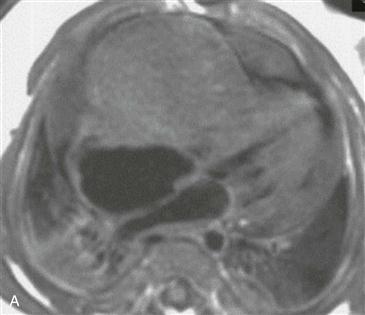
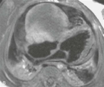
Cardiac fibroma is a rare, benign tumor; approximately 90% occur in children. Fibromas are usually well-marginated masses that are mainly composed of spindle cells and intervening collagen. Microscopic calcification is present in approximately half of these tumors.
MRI and Diagnostic Features
MRI demonstrates tumor size and location and cardiac function (Fig. A). Contrast-enhanced MRI can delineate tumor margins and extension and involvement of adjacent structures. However, the presence of enhancement is not reliable for determination of malignancy. Features that suggest a primary malignant cardiac tumor include invasiveness, extension outside the heart, involvement of more than one chamber, central necrosis or cavitation, and large pericardial effusion. Absence of these findings indicates benignancy. The appearance of fibroma is variable on spin echo T1-weighted images and cine MRI. Because fibroma is often isointense to myocardium, the administration of a contrast agent may be necessary to define the extent of a tumor. Fibromas have been reported to show irregular peripheral enhancement or heterogeneous enhancement with dark areas corresponding to calcification (Fig. B). Because gadolinium chelate contrast agent equilibrates rapidly into the extracellular space, the peripheral enhancement pattern indicates poor vascularization of the central fibrous tissue. The periphery of the mass is better vascularized and has a larger extracellular space. Because this enhancement pattern is comparable to that of a fast-growing tumor with central necrosis, it is not diagnostic of a fibroma. Definitive diagnosis requires endomyocardial or open biopsy, but MRI can be used to suggest the diagnosis of fibroma. Surgical resection is performed if the mass causes hemodynamic compromise.


