[level-membership-for-gastroenterology-and-hepatology-category]CASE 137
History: A 54-year-old man undergoes assessment for swelling of the left groin.
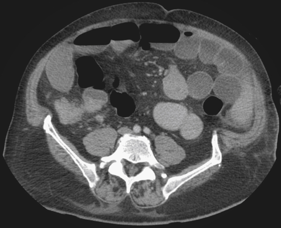
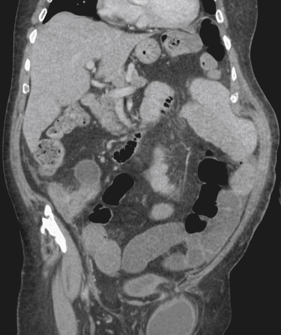
1. What should be included in the differential diagnosis of the imaging finding in the right lower quadrant shown in the figures? (Choose all that apply.)
2. What is the most common cause of an appendiceal mucocele?
D. Mucinous cystadenocarcinoma
3. What is the most common neoplasm of the appendix?
4. What is the most common abnormality seen on CT in appendicitis?
ANSWERS
CASE 137
Appendix Carcinoma
1. A, B, D, and E
2. C
3. D
4. D
References
Duran JC, Beidle TR, Perret R, et al: CT imaging of acute right lower quadrant disease. AJR Amer J Roentgenol. 1997;168(2):411–416.
Cross-Reference
Gastrointestinal Imaging: THE REQUISITES, 3rd ed, p 318.
Comment
Primary neoplasms of the appendix are uncommon, accounting for less than 1% of all gastrointestinal tumors. The most common are benign carcinoids. The most common primary malignant lesion of the appendix is adenocarcinoma, most frequently mucinous adenocarcinoma. Because of this and other mucoid lesions of the appendix, the presence of pseudomyxoma peritonei is always a possibility. Lesions are often found on routine appendectomy. Most carcinoids are found incidentally during postmortem examination.
The primary lesions are often small. At surgery, the appendix is found to be thickened and hard, and the leaves of the mesentery are studded with innumerable tiny yellow metastatic deposits. CT is the examination of choice (see figures). Although the disease is rare, if the thickened wall of the appendix does not look like edema, or there are adjacent nodes, hazy density of the mesentery, or evidence of distal disease, the diagnosis of neoplasia should be considered instead of the much more common diagnosis of appendicitis. Ascitic fluid is often present. An inflamed appendix may weep small amounts of fluid into the peritoneal cavity, which collects in the pelvic recesses; unless the ascites is significant, this is a less significant sign.
[/level-membership-for-gastroenterology-and-hepatology-category][not-level-membership-for-gastroenterology-and-hepatology-category]CASE 137
History: A 54-year-old man undergoes assessment for swelling of the left groin.
1. What should be included in the differential diagnosis of the imaging finding in the right lower quadrant shown in the figures? (Choose all that apply.)
2. What is the most common cause of an appendiceal mucocele?
D. Mucinous cystadenocarcinoma
3. What is the most common neoplasm of the appendix?
[/not-level-membership-for-gastroenterology-and-hepatology-category]
