CASE 136
History: An 83-year-old woman presents with suprasternal dysphagia.
1. What should be included in the differential diagnosis of the imaging finding shown in the figures? (Choose all that apply.)
D. Killian-Jamieson diverticulum
E. Lateral hypopharyngeal pouch
2. What is the anatomy of the site of weakness where a Killian-Jamieson diverticulum occurs?
B. Between the oblique and transverse fibers of the cricopharyngeus muscle
C. Between the inferior pharyngeal constrictor muscle and the thyrohyoid muscle
3. What specific complication is a patient at risk of with treatment of this lesion?
D. Injury of the recurrent laryngeal nerve
4. Which of the following is a true diverticulum?
C. Killian-Jamieson diverticulum
D. Intramural pseudodiverticulum
ANSWERS
CASE 136
Killian-Jamieson Diverticulum
1. B, C, D, and E
2. D
3. D
4. A
References
Ekberg O, Nylander G. Lateral diverticula from the pharyngo-esophageal junction area. Radiology. 1983;146(1):117–122.
Cross-Reference
Gastrointestinal Imaging: THE REQUISITES, 3rd ed, p 28.
Comment
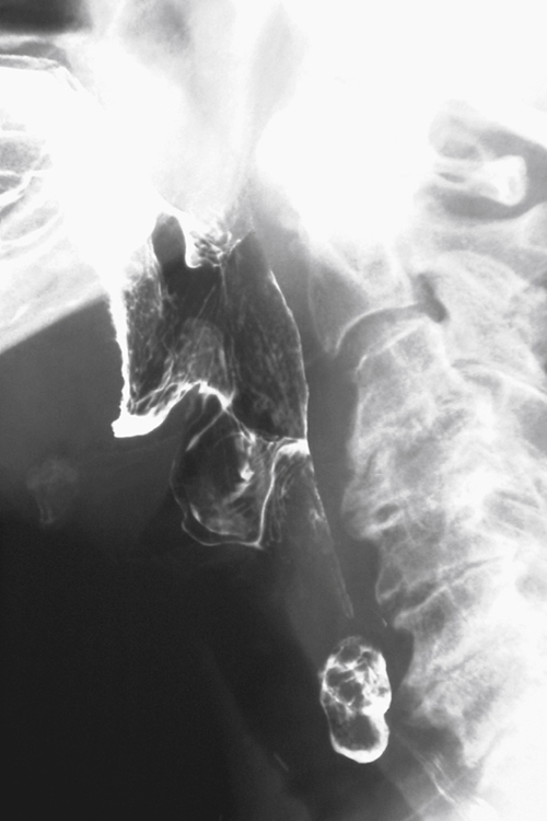
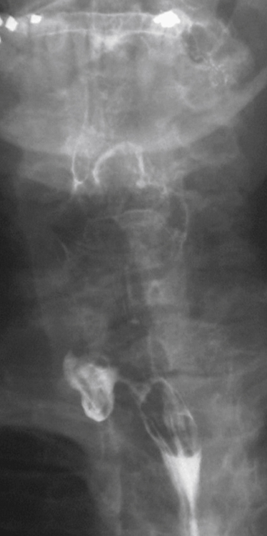
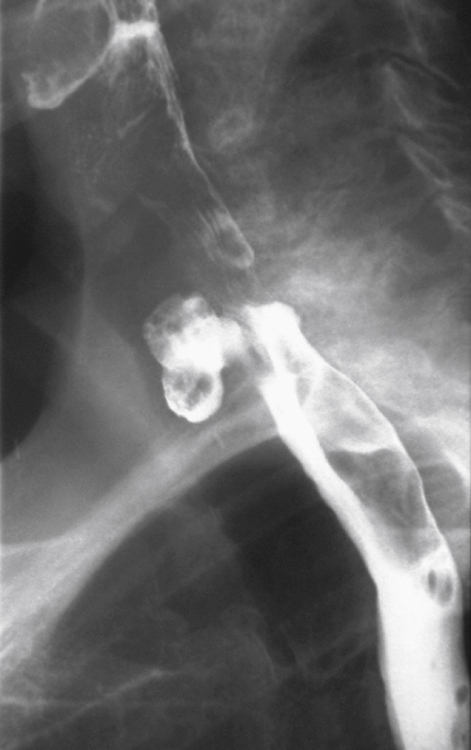
Also known as a proximal lateral cervical esophageal diverticulum, a Killian-Jamieson diverticulum is a pulsion diverticulum. It arises from an anatomic weak spot below the level of the cricopharyngeus muscle and lateral to the longitudinal muscles of the esophagus, just below its insertion into the cricoid cartilage.
Compared with the more common Zenker’s diverticulum, a Killian-Jamieson diverticulum is usually smaller in size and less likely to be symptomatic. Clinical distinction between the two types of diverticula is difficult because their symptoms are very similar. Radiologically, Zenker’s diverticulum can be confused with a Killian-Jamieson diverticulum (see figures). However, it is important to distinguish between the two because surgical management differs. A Killian-Jamieson diverticulum is in close proximity to the recurrent laryngeal nerve, and there is a tendency for surgical rather than endoscopic management of this diverticulum because of concerns regarding potential nerve injury.
Identification of the cricopharyngeus muscle is key to the diagnosis. The opening of a Zenker’s diverticulum lies above the protruding cricopharyngeus bar. The sac of a Zenker’s diverticulum is posterior to the cervical esophagus on lateral images and in the midline on frontal images. In contrast, the opening of a Killian-Jamieson diverticulum is below the cricopharyngeus bar. The sac of diverticulum overlaps the anterior wall of the cervical esophagus on lateral images and projects laterally on frontal images.


