[level-membership-for-gastroenterology-and-hepatology-category]CASE 135
History: A 45-year-old woman presents with fever, weight loss, lower abdominal pain, and an area of redness and swelling at the left iliac fossa.
1. What should be included in the differential diagnosis of the imaging finding shown in Figure A? (Choose all that apply.)
2. Which of the following statements regarding actinomycosis is true?
A. Penicillin is the treatment of choice.
B. The most common site of infection is in the chest.
C. The diagnosis is usually made by aspirate and culture.
D. Lymphatic and hematogenous spread readily occurs.
3. What is the most common site of actinomycosis of the gastrointestinal tract?
4. With what iatrogenic intervention is this infection in the pelvis associated?
A. Intrauterine contraceptive devices
ANSWERS
CASE 135
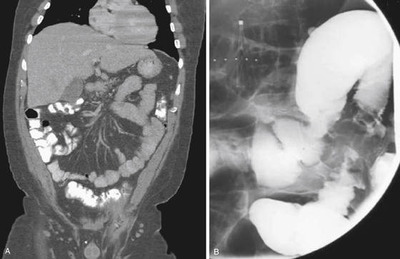
Actinomycosis of the Sigmoid Colon
1. A, B, C, and D
2. A
3. A
4. A
References
Ha HK, Lee HJ, Kim H, et al: Abdominal actinomycosis: CT findings in 10 patients. AJR Am J Roentgenol. 1993;161(4):791–794.
Cross-Reference
Gastrointestinal Imaging: THE REQUISITES, 3rd ed, p 317.
Comment
Actinomycosis is an uncommon inflammatory entity caused by the commonly found anaerobic bacterium, Actinomyces israelii, a gram-positive bacillus, which is a component of the normal human flora and can be found in the mouths of most people. When the cellular environment is conducive for anaerobic proliferation, it is possible for Actinomyces to proliferate, migrate, and infect tissue. It can occur almost anywhere in the body. Bowel involvement is uncommon, although it has increased in frequency over the last few decades. The most common site of the disease is the ileocolic junction. Involvement of the sigmoid colon has been described in patients with use of intrauterine contraceptive devices. Actinomycosis, similar to syphilis, can mimic other diseases, such as colonic diverticulitis, abscesses, appendagitis, and malignant tumors, presenting a diagnostic challenge and often being identified postoperatively in many cases.
The diagnosis can be determined with endoscopy and imaging techniques such as CT and MRI, where the striking characteristic of actinomycosis is best displayed: the tendency of the disease routinely to breach normal barriers to disease such as muscle and fascial planes (see figures). Other common findings in CT scan and barium study include mural invasion with stricture formation, mass effect with tapered narrowing of the lumen, and thickened mucosal folds (see figures). In many cases, the radiologic findings are similar to findings of intestinal tuberculosis and destructive malignant tumors.
[/level-membership-for-gastroenterology-and-hepatology-category][not-level-membership-for-gastroenterology-and-hepatology-category]CASE 135
History: A 45-year-old woman presents with fever, weight loss, lower abdominal pain, and an area of redness and swelling at the left iliac fossa.
1. What should be included in the differential diagnosis of the imaging finding shown in Figure A? (Choose all that apply.)
2. Which of the following statements regarding actinomycosis is true?
A. Penicillin is the treatment of choice.
B. The most common site of infection is in the chest.
C. The diagnosis is usually made by aspirate and culture.
D. Lymphatic and hematogenous spread readily occurs.
3. What is the most common site of actinomycosis of the gastrointestinal tract?
[/not-level-membership-for-gastroenterology-and-hepatology-category]
