CASE 135
1. What should be included in the differential diagnosis? (Choose all that apply.)
A. Aneurysm of right aortic arch
C. Right subclavian artery aneurysm
2. Which pharyngeal arch forms the normal aortic arch?
A. Third
B. Fourth
C. Fifth
D. Sixth
3. Which anomaly can be formed from the right third pharyngeal arch?
C. Left arch with aberrant right subclavian artery
4. Which of the following is the least appropriate next step in evaluation?
B. MRI
C. CT
ANSWERS
References
Caputo S, Villanacci R, Ciampi Q, et al. Cervical aortic arch: echocardiographic and three-dimensional computed tomography view. Echocardiography. 2010;27(4):E44–E45.
Poellinger A, Lembcke AE, Elgeti T, et al. Images in cardiovascular medicine The cervical aortic arch: a rare vascular anomaly. Circulation. 2008;117(20):2716–2717.
Cross-Reference
Cardiac Imaging: The REQUISITES, ed 3, p 427.
Comment
Etiology and Clinical Features
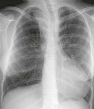
Cervical aortic arch is a rare anomaly in which the arch arises from the primitive third arch instead of the fourth. It may be more common on the right side. It has been reported that the ipsilateral internal and external carotid and vertebral arteries arise directly from the arch. Cervical aortic arch is usually asymptomatic but can manifest as a pulsatile mass in the supraclavicular fossa or neck, with obstruction secondary to kinking, or as an aneurysm.
Imaging and Diagnosis
Diagnosis is based on the presence of the aortic arch near the base of the neck (Figure). Some authors state that the diagnosis depends on the finding of separate origins of the internal and external carotid arteries directly from the arch. MRI and CT readily delineate the anatomy.


