CASE 132
History: A 43-year-old woman underwent a CT scan for renal colic, and findings suspicious for a mass in the cecum were incidentally noted.
1. What should be included in the differential diagnosis of the imaging finding shown in Figure A? (Choose all that apply.)
A. Fatty infiltration of the ileocecal valve
B. Lipoma of the ileocecal valve
2. Which of the following statements regarding the ileocecal valve at barium enema is true?
A. The ileocecal valve is usually directly visible.
B. A lobulated mucosal surface is suspicious for malignancy.
C. Asymmetry of the valve lips indicates infiltration by disease.
D. Filling of the terminal ileum is required to identify the ileocecal valve.
3. Which of the following statements regarding disease at the ileocecal valve is true?
C. The ileocecal valve is the most common site of development of cancer of the right colon.
D. A lipoma results in an asymmetric deformity of the ileocecal valve.
4. Which infective disease of the small bowel typically involves the terminal ileum?
ANSWERS
CASE 132
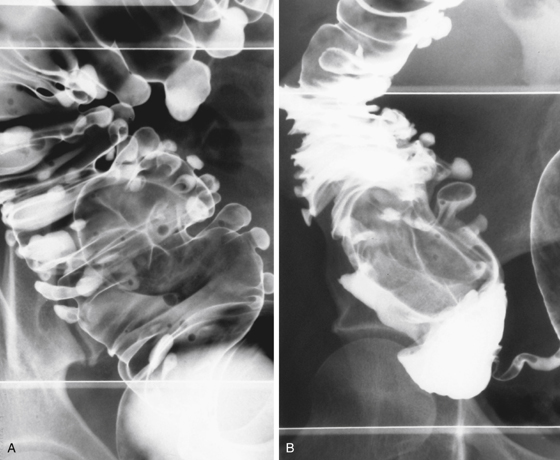
Fatty Infiltration of the Ileocecal Valve
1. A, C, and D
2. A
3. D
4. D
References
Dahnert W. Radiology Review Manual. 6th ed. Philadelphia: Lippincott Williams & Wilkins; 2007. p 780
Cross-Reference
Gastrointestinal Imaging: THE REQUISITES, 3rd ed, p 279.
Comment
With the development of CT, the opportunity to detect ileocecal disease has increased. Although a true ileocecal valve mass or cecal mass is the principal concern, other possibilities might warrant consideration. In this case, the “mass” proved to be fatty infiltration of the ileocecal valve, which is a benign condition.
A functional phenomenon of prolapse is sometimes the cause, which is visible with static imaging. It has long been suspected by fluoroscopists that prolapse through the ileocecal valve occurs when the right colon, and in particular, the ileocecal valve, is empty. Conversely, when the cecum becomes distended, the prolapsed ileum returns to its normal position, and there may be reflux of colonic material into the terminal ileum in some patients with incompetent ileocecal valves.
This condition must be distinguished from an ileocolonic intussusception, which is truly pathologic and almost always has an underlying cause and is invariably symptomatic. There are things to look for on CT studies of the abdomen: Evaluate the surrounding pericolic fat looking for stranding or small nodes. Evaluate the serosal wall of the bowel to ensure it is crisp. Make sure there is no suggestion of obstruction. If there is any concern, ask for a barium enema or dedicated colonography for confirmation (see figures).


