[level-membership-for-cardiovascular-category]
CASE 130
1. What should be included in the differential diagnosis? (Choose all that apply.)
C. Tetralogy of Fallot with absent pulmonary valve
2. Which vessel or vessels are enlarged?
A. Aorta
3. This patient has tetralogy of Fallot. What is the most likely diagnosis?
B. “Pink tet”
4. What is the cause of large lung volumes in these patients?
A. Emphysema
C. Infections
ANSWERS
Reference
Kirshbom PM, Kogon BE. Tetralogy of Fallot with absent pulmonary valve syndrome. Semin Thorac Cardiovasc Surg Pediatr Card Surg Annu. 2004;7:65–71.
Cross-Reference
Cardiac Imaging: The REQUISITES, ed 3, pp 359–367.
Comment
Overview
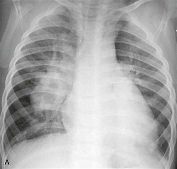
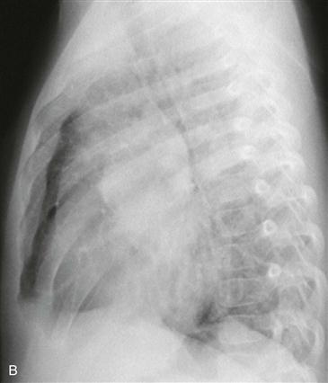
Tetralogy of Fallot is the most common cyanotic congenital heart disease in children and adults. The lesions of tetralogy of Fallot are ventricular septal defect, pulmonary infundibular stenosis, right ventricular hypertrophy, and overriding aorta. Most patients have decreased pulmonary vascularity, but vascularity can be normal if the infundibular stenosis is mild. Approximately 25% of patients have a right aortic arch. Pulmonary stenosis can occur at multiple levels, including subvalvular or infundibular (most common), valvular, supravalvular, and in peripheral pulmonary arteries. The chest radiograph typically demonstrates decreased vascularity, concavity of the pulmonary artery segment, and sometimes an upturned cardiac apex.
Clinical Features
Tetralogy of Fallot with absent pulmonary valve syndrome is characterized by a hypoplastic pulmonary annulus containing primitive valve tissue and aneurysmal dilation of the pulmonary arteries. The other lesions of tetralogy also exist in these patients. The marked pulmonary artery enlargement causes large airway compression, leading to early onset of respiratory distress.
[/level-membership-for-cardiovascular-category][not-level-membership-for-cardiovascular-category]
CASE 130
1. What should be included in the differential diagnosis? (Choose all that apply.)
C. Tetralogy of Fallot with absent pulmonary valve
2. Which vessel or vessels are enlarged?
A. Aorta
3. This patient has tetralogy of Fallot. What is the most likely diagnosis?
[/not-level-membership-for-cardiovascular-category]
