[level-membership-for-cardiovascular-category]
CASE 128

1. What should be included in the differential diagnosis for a dilated coronary artery? (Choose all that apply.)
B. Anomalous left coronary artery from the pulmonary artery
C. Fistula
2. Which coronary artery is enlarged?
A. Right
B. Left main
3. Which structure is opacified at the same time as the left circumflex coronary artery at the inferior aspect of the heart (Fig. C)?
4. What is the abnormality affecting the left circumflex coronary artery?
A. Dissection
B. Aneurysm
C. Fistula
ANSWERS
Reference
Díaz-Zamudio M, Bacilio-Pérez U, Herrera-Zarza MC, et al. Coronary artery aneurysms and ectasia: role of coronary CT angiography. Radiographics. 2009;29(7):1939–1954.
Cross-Reference
Cardiac Imaging: The REQUISITES, ed 3, pp 228–229, 253.
Comment
Imaging
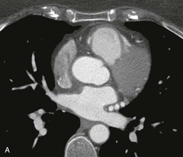
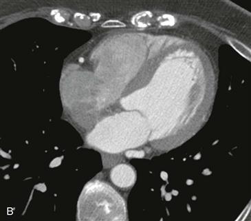
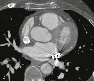
Three axial images from CT angiography of the coronary arteries show marked dilation and tortuosity of the circumflex coronary artery (Figs. A–C). There is contrast opacification of the distal coronary sinus (Fig. C) without opacification of the more proximal great cardiac vein; this is consistent with a coronary artery fistula to the coronary sinus. Syncope in this patient was thought to be secondary to a coronary steal phenomenon that occurred with exercise, and she was treated successfully with coil embolization of the distal circumflex coronary artery (Fig. D).
Description
A coronary fistula is usually a congenital connection between a coronary artery and cardiac chamber or great vessel that bypasses the myocardial capillaries. Coronary fistulas can rarely occur after myocardial biopsy or open heart surgery. The right coronary artery is affected in most cases (52%), followed by the left anterior descending coronary artery (30%) and the left circumflex coronary artery (18%). In most patients (90%), the fistula drains into the right circulation causing a left-to-right shunt.
[/level-membership-for-cardiovascular-category][not-level-membership-for-cardiovascular-category]
CASE 128
1. What should be included in the differential diagnosis for a dilated coronary artery? (Choose all that apply.)
B. Anomalous left coronary artery from the pulmonary artery
C. Fistula
2. Which coronary artery is enlarged?
A. Right
B. Left main
[/not-level-membership-for-cardiovascular-category]
