CASE 126
1. What are causes of right heart failure? (Choose all that apply.)
B. Emphysema
C. Idiopathic pulmonary fibrosis
E. Congenital pulmonary stenosis
2. What is the main finding involving the cardiac chambers?
A. Dilation of the right-sided chambers
B. Dilation of left-sided chambers
3. What valve abnormality would explain the dilated chambers?
ANSWERS
Reference
Yalonetsky S, Tobler D, Greutmann M, et al. Cardiac magnetic resonance imaging and the assessment of Ebstein anomaly in adults. Am J Cardiol. 2011;107(5):767–773.
Cross-Reference
Cardiac Imaging: The REQUISITES, ed 3, pp 206–207.
Comment
Description
Ebstein anomaly is a rare congenital heart disease that usually results in cyanosis early in life. This anomaly is characterized by varying degrees of ventricular displacement of the tricuspid valve leaflets, resulting in a common ventriculoatrial chamber and often severe tricuspid regurgitation. The tricuspid anulus is not displaced. Over time, there is progressive dilation of the right-sided chambers, which leads to increased tricuspid regurgitation and further chamber dilation. Occasionally, the degree of ventricular displacement of the tricuspid leaflets is mild, resulting in less tricuspid regurgitation and less right atrial dilation. These patients commonly present later in life and sometimes as adults.
Imaging
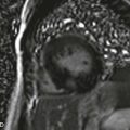
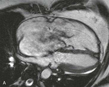
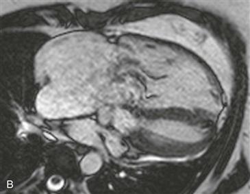
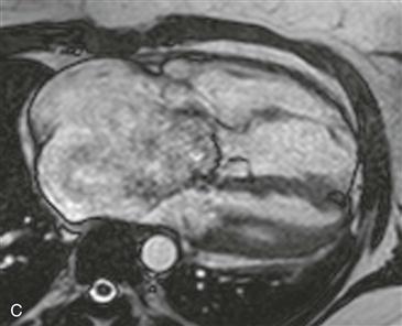
Cine steady-state free precession (SSFP) images (Figs. A–C) in the four-chamber view show dilation of the right atrium and right ventricle. The tricuspid valve leaflets are displaced toward the right ventricle, and there is associated severe tricuspid regurgitation, with the tricuspid regurgitant fraction measuring 74%. This patient had a late presentation of Ebstein anomaly. She noted fatigue beginning in her early 20s, which progressed to exercise intolerance and lower extremity edema.
Role of MRI in Adults
Echocardiography allows precise morphologic and functional evaluation of the right ventricle in children with Ebstein anomaly. This evaluation is important because right ventricular size is directly correlated to patient outcomes and prognosis. In adults, the acoustic windows needed to image the right atrium and ventricle on echocardiography may be inadequate or allow incomplete visualization of the chambers. MRI provides the same information as echocardiography and gives more reliable and reproducible information in patients with limited acoustic windows. MRI can assess right ventricular size and function and accurately quantitate the volume of tricuspid regurgitation, which has a direct impact on the timing of valvular surgery in these patients.