CASE 122
History: A 64-year-old woman presents with nausea and vomiting.
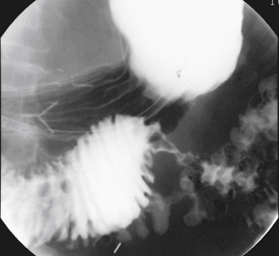
1. What should be included in the differential diagnosis of the imaging finding shown in the figure? (Choose all that apply.)
2. What is the most common primary malignancy of the duodenum?
3. What is the most common location of duodenal carcinoma?
4. Which of the following diseases places a patient at greatest risk of developing duodenal carcinoma?
ANSWERS
CASE 122
Duodenal Primary Cancer
1. A, B, C, and E
2. C
3. B
4. D
References
Bradford D, Levine MS, Hoang D, et al: Early duodenal cancer: detection on double-contrast upper gastrointestinal radiography. Am J Roentgenol. 2000;174(6):1564–1566.
Cross-Reference
Gastrointestinal Imaging: THE REQUISITES, 3rd ed, p 91.
Comment
Primary adenocarcinoma, such as shown in this case (see figure), is uncommon. It is thought to represent 0.3% of all malignancies of the GI tract. Most patients are slow to seek medical attention because the lesion is insidious and the symptoms are nonspecific, and it is often fatal by the time of diagnosis. In one study, the diagnosis was made after death in 25% of cases. At least 50% of patients have metastatic disease at the time of diagnosis. With the increased use of widespread endoscopy and abdominal CT examinations, this percentage should have improved in the last 15 years. However, whether increased use of these imaging examinations has affected long-term survival rates is still unclear. The incidence of primary adenocarcinoma of the duodenum is increased in patients with Gardner’s familial polyposis, as is the incidence of adenoma. The more proximal the lesion is located, the earlier a diagnosis is made, and survival is improved. These patients may also present with biliary symptoms.


