CASE 120
History: A 38-year-old woman presents with diarrhea.
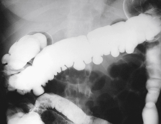
1. Which of the following should be included in the differential diagnosis of the imaging finding shown in the figure? (Choose all that apply.)
2. Which of the following lesions of the gastrointestinal tract are “true” diverticula?
A. Sacculations of scleroderma
3. What is the most common gastrointestinal tract site of involvement by scleroderma?
4. Which of the following is the least common complication of colonic scleroderma?
ANSWERS
CASE 120
Colonic Scleroderma
1. A, C, D, and E
2. A
3. A
4. A
References
Cohen S, Laufer I, Snape WJ Jr., et al: The gastrointestinal manifestations of scleroderma: pathogenesis and management. Gastroenterology. 1980;79:155–166.
Cross-Reference
Gastrointestinal Imaging: THE REQUISITES, 3rd ed, p 312.
Comment
The presence of large wide-mouth sacculations in the colon can be an indication of several diseases (see figure). Many term these wide-mouth diverticula pseudodiverticula because they are not related to the typical diverticula seen in everyday practice. However, they contain all three layers of bowel and thus represent true diverticula. The small diverticula seen in everyday practice are not true diverticula. These large-mouth diverticula or sacculations occur when there is eccentric involvement of the bowel wall by some process, producing fibrosis on one side, with eccentric bulging of the opposite wall.
A variety of processes can result in the loss of the haustral folds, but only some produce the eccentric diverticula. These processes include scleroderma, Crohn’s disease, and laxative abuse. Scleroderma does not involve the colon as commonly as it does other portions of the gastrointestinal tract. Just as it does in other portions of the gastrointestinal tract, however, scleroderma of the colon produces patchy smooth muscle atrophy along with fibrotic replacement of the muscle. This effect leads to the formation of wide-mouth sacculations at weakened areas. There are also abnormalities of transit time, and patients often complain of constipation. The haustral fold pattern is often diminished. Fecal impaction is a complication, and patients can develop benign pneumatosis, which could be the result of a combination of steroid therapy and stasis in the colon.
An interesting feature of these wide-mouth sacculations is their ability to retain material. They fail to contract and empty and are found outside the fecal stream in the colon. Thus, develop impacted fecal material or fecaliths sometimes develop within sacculations. These fecaliths can become adherent and resemble polyps or tumors on barium enema studies. Although scleroderma can result in colonic sacculations, the classic hidebound appearance of scleroderma is limited to small bowel involvement. Fibrosis in the wall and folds of the small bowel give rise to this unusual appearance of atrophic folds, seemingly pulled tightly together by the disease. This should not be confused with the “stack of coins” appearance of small bowel intramural hemorrhage in which the folds show no effacement.


