CASE 120
1. What should be included in the differential diagnosis for a saccular outpouching from a vessel? (Choose all that apply.)
2. What is the most likely diagnosis?
3. Where is the most common site of rupture of this entity?
A. Pericardium
4. What is the most common complication from rupture?
A. Hemothorax
ANSWERS
Reference
Bricker AO, Avutu B, Mohammed TL, et al. Valsalva sinus aneurysms: findings at CT and MR imaging. Radiographics. 2010;30(1):99–110.
Cross-Reference
Cardiac Imaging: The REQUISITES, ed 3, pp 389–392.
Comment
Epidemiology
Sinus of Valsalva aneurysms are rare congenital or acquired lesions that occur in 0.09% of patients as determined in a large autopsy study. They are more common in men and people of Asian descent. The right coronary cusp is affected in most cases (72%) followed by the noncoronary cusp (22%). When symptoms exist, they are often secondary to rupture or mass effect on a coronary artery, superior vena cava, or ventricular outflow tract. Sinus of Valsalva aneurysms most commonly rupture into the right ventricle followed by the right atrium. Rupture into a cardiac chamber causes an aortocardiac shunt with the eventual development of congestive heart failure. Large nonruptured aneurysms may cause mass effect on adjacent structures or lead to aortic regurgitation.
Diagnosis and Treatment
Sinus of Valsalva aneurysms are most commonly diagnosed with echocardiography. Occasionally, CT or MRI may be used in difficult cases, or aneurysms may be discovered on imaging performed for other reasons. The imaging criteria to diagnose this condition include a saccular shape, aneurysm origin above the aortic anulus, and a normal dimension of the aortic root and ascending aorta. Definitive treatment is cardiopulmonary bypass surgery or percutaneous repair.
Imaging
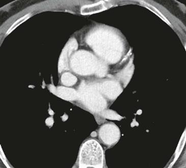
Contrast-enhanced CT through the level of the aortic root shows a saccular outpouching arising from the left sinus of Valsalva (Figure). The adjacent aortic root and ascending aorta have a normal diameter. No communication with the ventricle is present. These findings are consistent with an unruptured left coronary sinus of Valsalva aneurysm.


