CASE 12
1. Which of the following are risk factors for this diagnosis? (Choose all that apply.)
B. Malignancy
C. Paralysis
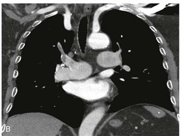
2. What cardiac finding is seen in Fig. B?
A. Leftward bowing of interventricular septum
3. What is the most likely cause of the cardiac finding in this patient with pulmonary embolism?
B. Normal
4. Which of these findings indicates right heart strain in a patient with pulmonary embolism?
A. Rightward bowing of interventricular septum
B. Main pulmonary artery diameter greater than 20 mm
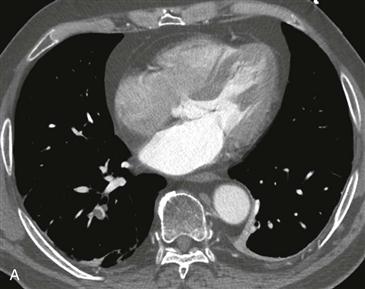
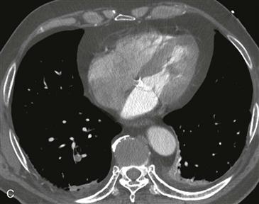
C. Right ventricular-to-left ventricular (RV/LV) ratio greater than 1
ANSWERS
Reference
Ghaye B, Ghuysen A, Bruyere PJ, et al. Can CT pulmonary angiography allow assessment of severity and prognosis in patients presenting with pulmonary embolism? What the radiologist needs to know. Radiographics. 2006;26(1):23–39. discussion 39–40.
Comment
Imaging
Axial and coronal contrast-enhanced CT pulmonary angiogram images (Figs. A–C) show multiple segmental and right upper lobar pulmonary emboli. Images of the heart show leftward bowing of the interventricular septum and an elevated RV/LV ratio measuring 1.2. This elevated ratio indicates right heart strain.
Prognostic Indicators
Pulmonary embolism remains a common clinical problem despite advances in thromboembolism prophylaxis. Two large multicenter trials found a significantly higher mortality in patients presenting with hemodynamic instability (50% to 58% mortality compared with 8% to 15% mortality in hemodynamically stable patients). Several CT findings have been shown to be predictive of right heart strain and impending hemodynamic instability. Commenting on these CT findings in an imaging report allows the clinician to initiate appropriate management and patient monitoring. The most sensitive and specific imaging sign is an abnormal RV/LV ratio greater than 1 (Figs. A and C). This ratio compares the short-axis diameters of the right ventricle and left ventricle. The right ventricular and left ventricular short axis diameters are measured on axial images at the respective levels of the tricuspid and mitral valves. The largest inner-to-inner diameter of the right ventricle and left ventricle is taken to measure the short-axis diameter. In normal patients, the left ventricular short axis is always larger than the right ventricular short axis, and the RV/LV ratio is less than 1. A RV/LV ratio greater than 1 is a highly sensitive and specific sign of right ventricular strain. An abnormal RV/LV ratio is predictive of higher patient mortality and a 3.6 times higher rate of intensive care unit admission. Other signs of right heart strain include leftward interventricular septal shift and a main pulmonary arterial diameter greater than 30 mm.


