CASE 119
1. What should be included in the differential diagnosis for myocardial late gadolinium enhancement? (Choose all that apply.)
D. Hypertrophic cardiomyopathy
2. What MRI sequence is shown in Figs. A and B?
C. Steady-state free precession (SSFP) MRI
D. Inversion recovery late gadolinium enhancement MRI
3. What is the most likely cause of the hyperintense signal within the right ventricular wall?
C. Amyloidosis
D. Hypertrophic cardiomyopathy
4. Which cardiac MRI sequence can identify microvascular obstruction?
A. Cine steady-state free precession
B. Velocity-encoded cine phase contrast MRI
ANSWERS
References
Ito H. No-reflow phenomenon and prognosis in patients with acute myocardial infarction. Nat Clin Pract Cardiovasc Med. 2006;3(9):499–506.
Mather AN, Lockie T, Nagel E, et al. Appearance of microvascular obstruction on high resolution first-pass perfusion, early and late gadolinium enhancement CMR in patients with acute myocardial infarction. J Cardiovasc Magn Reson. 2009;11:33.
Comment
Imaging
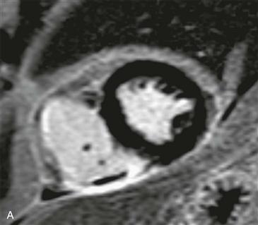
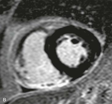
No-reflow zone can be detected on first-pass perfusion imaging and early and late gadolinium imaging. Because contrast material cannot reach the myocardium owing to microvascular obstruction, it remains dark on all postgadolinium sequences (Figs. A and B). First-pass perfusion imaging is the most accurate predictor of the size of a no-reflow zone. Late gadolinium images likely underestimate the degree of microvascular obstruction because contrast material often diffuses into the edge of the no-reflow zone.
Pathophysiology and Prognosis
Cardiac MRI is a useful prognostic tool after intervention for acute ST segment elevation myocardial infarction because it directly quantifies the volume of infarcted myocardium and the extent of microvascular obstruction. After myocardial infarction, no-reflow zone is caused by a breakdown or obstruction of the coronary microvasculature in the territory supplied by the previously obstructed coronary artery. The presence of a no-reflow zone is a poor prognostic indicator; patients have an increased future risk of lower ejection fraction, more ventricular remodeling, and higher risk of death.


