[level-membership-for-gastroenterology-and-hepatology-category]CASE 117
History: A 75-year-old man presents with bloody diarrhea.
1. Which of the following should be included in the differential diagnosis of the imaging finding shown in the figure? (Choose all that apply.)
2. Which of the following statements regarding colonic amebiasis is true?
A. Usually involves the left colon
B. Typically involves the terminal ileum
C. Usually appears as a diffuse ulcerative colitis
D. Sometimes occurs as a short masslike narrow segment
3. What is the typical change produced by ulcerative colitis in the terminal ileum?
A. Dilated gaping terminal ileum
B. Narrowing, nodular mucosa, fissures, and fistulas
C. Asymmetrical luminal narrowing with spiculated contour
D. Mucosal nodularity with fold thickening and superficial ulceration
4. What fungal infection can produce this abnormality?
ANSWERS
CASE 117
Colonic Amebiasis
1. A, B, C, and D
2. D
3. A
4. B
References
Cardoso JM, Kimura K, Stoopen M, et al: Radiology of invasive amebiasis of the colon. Am J Roentgenol. 1977;128:935–941.
Cross-Reference
Gastrointestinal Imaging: THE REQUISITES, 3rd ed, p 299.
Comment
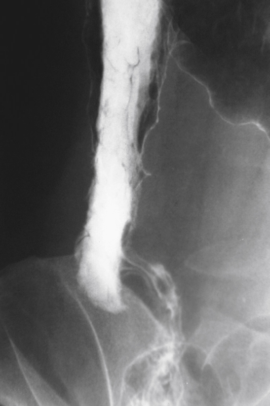
The coned appearance of the cecum is produced by several diseases, most of which are inflammatory in nature. In determining the exact cause, the radiologist must make a proper evaluation of the region. The most common causative condition encountered in industrialized societies is Crohn’s disease. Tuberculosis can manifest with an appearance virtually identical to that of Crohn’s disease, with a coned cecum and inflammation of the terminal ileum. Adjacent inflammatory conditions, such as appendicitis and diverticulitis, are considerations.
Amebiasis is an infection of the bowel produced by the protozoan Entamoeba histolytica. It is acquired by ingestion of water or soil that contains the cysts. When infection occurs, it can range from very mild or indolent to severe, acute colitis. When cysts spread to the liver or lungs, they can produce abscesses. Changes in the colon include ulceration, which can be either diffuse granularity, collar-button ulcers, or aphthous ulcers. The colon can have skip lesions, with intervening areas of normal bowel, and thus can resemble Crohn’s disease. Focal severe inflammation can mimic annular carcinomas. There also may be pronounced granulation tissue, leading to protuberant lesions called amebomas, which can also mimic neoplasia.
The cecum is invariably infected in amebiasis, and the classic appearance is that of the coned cecum (see figure). This abnormality is often seen in the chronic stages of colitis. One strong differential consideration is that amebiasis does not affect the terminal ileum, as do Crohn’s disease and tuberculosis. However, amebiasis is seen predominantly in underdeveloped countries and should be considered only if the history is appropriate.
Neoplasms, such as adenocarcinoma and lymphoma, are also in the differential diagnosis. Long-standing ulcerative colitis can produce a coned cecum but often with a dilated terminal ileum, called backwash ileitis. Other rare conditions that can produce this appearance include anisakiasis, blastomycosis, and infection by Yersinia species.
[/level-membership-for-gastroenterology-and-hepatology-category][not-level-membership-for-gastroenterology-and-hepatology-category]CASE 117
History: A 75-year-old man presents with bloody diarrhea.
1. Which of the following should be included in the differential diagnosis of the imaging finding shown in the figure? (Choose all that apply.)
2. Which of the following statements regarding colonic amebiasis is true?
A. Usually involves the left colon
B. Typically involves the terminal ileum
C. Usually appears as a diffuse ulcerative colitis
D. Sometimes occurs as a short masslike narrow segment
3. What is the typical change produced by ulcerative colitis in the terminal ileum?
A. Dilated gaping terminal ileum
B. Narrowing, nodular mucosa, fissures, and fistulas
C. Asymmetrical luminal narrowing with spiculated contour
D. Mucosal nodularity with fold thickening and superficial ulceration
[/not-level-membership-for-gastroenterology-and-hepatology-category]
