CASE 117
1. Which findings are present? (Choose all that apply.)
B. Cardiomegaly
C. Deviation of trachea and esophagus
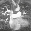
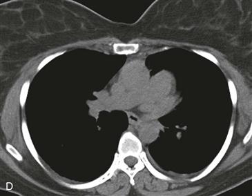
D. Lung between the aorta and pulmonary artery
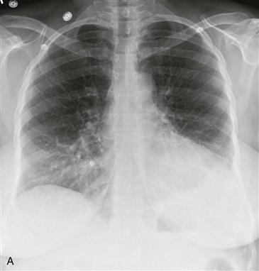
2. Which structure is bulging along the left heart border?
A. Aorta
3. What is the most likely diagnosis?
4. What is the most appropriate management?
A. No further evaluation or treatment
D. Surgery
ANSWERS
Reference
Wang ZJ, Reddy GP, Gotway MB, et al. CT and MR imaging of pericardial disease. Radiographics. 2003;23(Spec No):S167–S180.
Cross-Reference
Cardiac Imaging: The REQUISITES, ed 3, p 78.
Comment
Clinical Features
Congenital absence of the left pericardium is usually asymptomatic and needs no treatment. It is thought to occur secondary to a compromise of vascular supply to the pericardium during embryogenesis. If the pericardial defect is small and the left atrium, atrial appendage, or left ventricle herniates through it and becomes strangulated, the patient may experience chest pain, syncope, or sudden death. Surgery may be required in cases of cardiac herniation to close or enlarge the pericardial defect.
Types and Associations
The most common type is congenital absence of the left pericardium with preservation of the right pericardium (as in the case shown). Occasionally, congenital absence of the pericardium is associated with other congenital heart defects, including atrial septal defect, patent ductus arteriosus, mitral valve stenosis, and tetralogy of Fallot.
Imaging Findings and Diagnosis
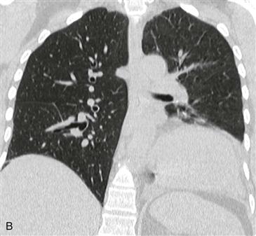
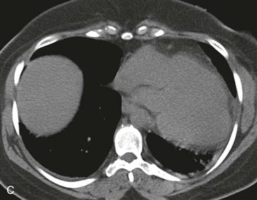
Chest radiographs typically demonstrate the characteristic leftward deviation and rotation of the heart without deviation of the remainder of the mediastinum and prominence of the left atrial appendage (Fig. A). These features are also seen on CT and MRI (Figs. B and C). Demonstration of lung interposed between the aorta and pulmonary artery on CT or MRI confirms the diagnosis (Fig. D).