CASE 114
1. What should be included in the differential diagnosis? (Choose all that apply.)
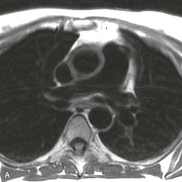
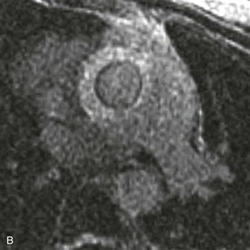
2. Which artery has a thickened, enhancing wall?
3. What is the most likely diagnosis?
4. What artery is most commonly involved in this disease?
A. Aorta
ANSWERS
References
Gotway MB, Araoz PA, Macedo TA, et al. Imaging findings in Takayasu’s arteritis. AJR Am J Roentgenol. 2005;184(6):1945–1950.
Reddy GP, Gunn M, Mitsumori LM, et al. Multislice CT and MRI of the thoracic aorta. In: Webb WR, Higgins CB, eds. Thoracic imaging: pulmonary and cardiovascular radiology. ed 2 Philadelphia: Lippincott Williams & Wilkins; 2010.
Cross-Reference
Cardiac Imaging: The REQUISITES, ed 3, pp 393–394.
Comment
Clinical Features
Takayasu arteritis (synonyms include pulseless disease and Martorell syndrome) is an idiopathic disease that is characterized by wall thickening of the aorta or arch vessels (or both) with stenosis of the aorta and its branches. Other arteries, such as the pulmonary arteries, can be involved. The two main large vessel arteritides include Takayasu arteritis and giant cell arteritis. Takayasu arteritis most commonly affects Asian women 10 to 40 years of age, whereas giant cell arteritis occurs in patients older than 50. The treatment of choice generally is high-dose corticosteroids.
Imaging
In Takayasu arteritis, MRI and CT can show stenosis, occlusion, or dilation of the aorta and its branches, or a combination of all three. In patients in the active phase of arteritis, gadolinium-enhanced MRI demonstrates wall thickening and enhancement of the involved vessels (Figs. A and B). Noninvasive imaging with MRI or CT may be particularly valuable in patients with severe stenosis or occlusion of the arch vessels or of the abdominal aorta because it may be especially difficult to pass an angiography catheter into the thoracic aorta. An aortic wall thickness of greater than 3 mm has been proposed as a marker for early Takayasu arteritis. Concentric aortic wall calcification has been described in patients with late-stage Takayasu arteritis. Positron emission tomography (PET) is useful for monitoring disease response in patients with large vessel vasculitides. Decreasing fluorodeoxyglucose (FDG) activity is considered a favorable response to therapy and may be seen before decreased aortic wall thickening is shown on anatomic imaging.


