CASE 112
1. What are advantages of CT over MRI for evaluation of the thoracic aorta? (Choose all that apply.)
A. No need for electrocardiogram (ECG) gating
B. No need for gadolinium-based contrast agent
2. Which of the following is present in pseudocoarctation?
B. Hemodynamically significant narrowing
D. Rib notching
3. What is the most likely diagnosis?
A. Coarctation
ANSWERS
References
Reddy GP, Gunn M, Mitsumori LM, et al. Multislice CT and MRI of the thoracic aorta. In: Webb WR, Higgins CB, eds. Thoracic imaging: pulmonary and cardiovascular radiology. ed 2 Philadelphia: Lippincott Williams & Wilkins; 2010.
Sebastia C, Quiroga S, Boye R, et al. Aortic stenosis: spectrum of diseases depicted at multisection CT. Radiographics. 2003;23(Spec No):S79–S91.
Cross-Reference
Cardiac Imaging: The REQUISITES, ed 3, pp 424–427.
Comment
Etiology and Anatomy
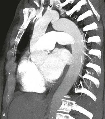
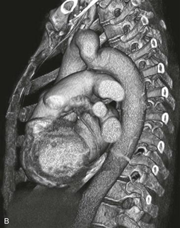
Pseudocoarctation is an uncommon congenital anomaly caused by elongation of the aortic arch and kinking or buckling at the site of attachment to the ligamentum arteriosum. There is no narrowing of the vessel lumen, and there is no hemodynamic gradient. Similar to aortic coarctation, there is an association with a congenitally bicuspid aortic valve, which should be excluded with echocardiography. Patients are generally asymptomatic, and the anomaly is discovered on imaging performed for other reasons. No treatment is necessary in most cases.
Imaging
Radiographs are often nonspecific, but they can show an abnormal aortic knob and a “figure 3” contour of the proximal descending aorta. On CT and MRI, the appearance of the aorta in pseudocoarctation is similar to aorta in true coarctation (Figs. A and B). CT has the disadvantage of ionizing radiation, and MRI may be better at distinguishing pseudocoarctation from aortic coarctation. MRI determines functional information by measuring the pressure gradient across the narrowing and by determining the volume of collateral flow through the use of velocity-encoded cine phase contrast MRI. In pseudocoarctation, there is no narrowing of the vessel lumen and no pressure gradient across the site of kinking. Collateral circulation does not develop, and rib notching does not occur. Thoracic aortic aneurysms occasionally develop around the pseudocoarctation, and these should be monitored closely and treated.


