CASE 111
1. Which of the following are advantages of MRI over CT for the evaluation of congenital heart disease? (Choose all that apply.)
A. MRI is safer in patients with renal insufficiency.
B. MRI can determine function.
C. MRI provides better spatial resolution.
D. MRI has a lower radiation dose.
2. What is the most likely diagnosis?
A. Aortic arch anomaly or vascular ring
3. In infancy, which of the following symptoms or complications is least likely?
B. Murmur
D. Cyanosis
4. Which of the following is not an appropriate treatment in this patient?
A. Intravenous indomethacin or ibuprofen
ANSWERS
References
Kilner PJ. Imaging congenital heart disease in adults. Br J Radiol. 2011;84(Spec No 3):S258–S268.
Wang ZJ, Reddy GP, Gotway MB, et al. Cardiovascular shunts: MR imaging evaluation. Radiographics. 2003;23(Spec No):S181–S194.
Cross-Reference
Cardiac Imaging: The REQUISITES, ed 3, pp 345–349.
Comment
Embryology, Clinical Features, and Treatment
In the fetus, the ductus arteriosus is a widely patent vessel that connects the proximal descending aorta to the main or left pulmonary artery, allowing the output of the right ventricle to bypass the lungs. The ductus usually closes shortly after birth as a result of the increase in the partial pressure of oxygen in the circulation. If the ductus does not close, intravenous indomethacin or ibuprofen can be administered in the first week of life, especially in premature infants. If the ductus remains patent, pulmonary hypertension can develop. In the absence of a coexisting anomaly, the ductus should be closed by surgical ligation or by transcatheter coil embolization or occluder device.
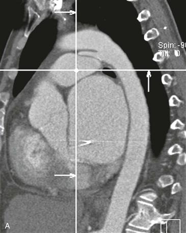
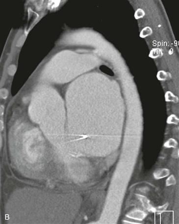
Imaging
Radiography can demonstrate increased pulmonary vascularity and enlargement of the left atrium and aortic arch. Echocardiography usually depicts the location and size of the lesion. MRI or CT may be performed in certain patients for better delineation of the lesion (Figs. A and B), and velocity-encoded cine phase contrast MRI can be performed for quantification of the pulmonary-to-systemic flow ratio, which is an indicator of the severity of the shunt.


