CASE 110
1. What etiologies can cause the cardiac finding? (Choose all that apply.)
A. Tuberculosis
B. Uremia
C. Mesothelioma
D. Hemorrhage
2. Which cardiac structure is calcified?
B. Pericardium
C. Valve
3. Given the patient’s clinical presentation, what is the most likely diagnosis?
A. Tumor
B. Tamponade
4. Given the other findings in the chest, what is the most likely etiology of the cardiac abnormality?
A. Tuberculosis
B. Uremia
C. Mesothelioma
D. Hemorrhage
ANSWERS
References
Gowda RM, Boxt LM. Calcifications of the heart. Radiol Clin North Am. 2004;42(3):603–617.
Walker CM, Reddy GP, Steiner RM. Radiology of the heart. In: Rosendorff C, ed. Essential Cardiology. ed 3 Boston: Saunders; 2012.
Cross-Reference
Cardiac Imaging: The REQUISITES, ed 3, pp 10, 79–82.
Comment
Calcific Pericarditis
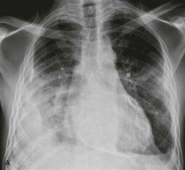
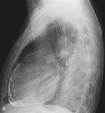
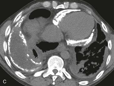
Chronic pericarditis can follow uremic pericarditis, viral or tuberculous infection, radiation therapy, or open heart surgery. In chronic pericarditis, pericardial calcification most often results from tuberculosis. Because tuberculous pericarditis is rare in industrialized countries, pericardial calcification (Figs. A–C) occurs in less than 20% of patients with chronic pericarditis. Calcific pericarditis does not necessarily cause symptoms of constriction.
Constrictive Pericarditis
In the setting of chronic pericarditis, a patient may have constrictive pericarditis. Constrictive pericarditis is difficult to distinguish from restrictive cardiomyopathy. If a patient has constrictive/restrictive physiology, characterized by dyspnea, lower extremity edema, pleural effusions, and ascites, imaging studies can be used to differentiate constrictive pericarditis from restrictive cardiomyopathy. This differentiation is important because treatments are vastly different. Constrictive pericarditis is treated surgically with pericardial stripping, whereas restrictive cardiomyopathy is managed medically or with heart transplantation. Pericardial thickening of at least 4 mm (best seen on MRI or CT), pericardial calcification (Figs. A–C), and abnormal diastolic septal motion (“septal bounce”) help establish the diagnosis of constrictive pericarditis.
Restrictive Cardiomyopathy
Patients with restrictive cardiomyopathy and patients with constrictive pericarditis have similar symptoms. On imaging, the pericardial thickness and systolic function are generally normal. Symptoms are due to diastolic dysfunction leading to restricted ventricular filling and reduced cardiac output. Inversion recovery late gadolinium enhancement imaging is helpful in suggesting a specific diagnosis. Causes of restrictive cardiomyopathy include amyloidosis, radiation therapy, sarcoidosis, and hemochromatosis.


