[level-membership-for-cardiovascular-category]
CASE 108
1. What should be included in the differential diagnosis? (Choose all that apply.)
A. Coarctation
2. The patient’s blood pressure is unknown. If the patient has normal systemic blood pressure, what is the most likely diagnosis, based on the radiograph?
A. Coarctation
3. Based on the CT image, what is the most likely diagnosis?
A. Coarctation
4. What is an advantage of CT over MRI for evaluation of coarctation of the aorta?
A. It is safer in patients with renal insufficiency.
B. It can determine functional significance.
ANSWERS
References
Hom JJ, Ordovas K, Reddy GP. Velocity-encoded cine MR imaging in aortic coarctation: functional assessment of hemodynamic events. Radiographics. 2008;28(2):407–416.
Kilner PJ. Imaging congenital heart disease in adults. Br J Radiol. 2011;84(Spec No 3):S258–S268.
Cross-Reference
Cardiac Imaging: The REQUISITES, ed 3, pp 72, 160–162, 420.
Comment
Clinical Features
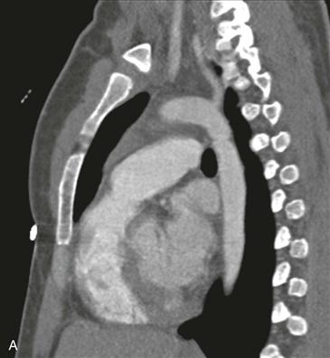
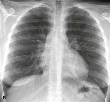
Congenital coarctation is most commonly discrete and juxtaductal in location (Fig. A). Hypertension is a common finding, often localized to the upper extremities.
Imaging
Radiographs may be nonspecific, but they can show an abnormal aortic knob (Fig. B), a “figure 3” contour of the proximal descending aorta, and rib notching. Rib notching occurs as a result of dilated intercostal arteries, which function as a collateral route for blood flow around the site of aortic narrowing. The main pathway of collateral flow proceeds as follows: aortic arch to subclavian artery to internal mammary artery to intercostal artery to descending thoracic aorta. Additional collateral pathways around the coarctation include the thyrocervical, thoracoacromial trunk, and epigastric arteries. Echocardiography can demonstrate the pressure gradient across the stenosis. CT and MRI can be used to delineate the anatomy. CT has the disadvantage of ionizing radiation, and MRI has the ability to determine the hemodynamic significance of the coarctation. Another benefit of MRI over CT and other modalities is its ability to distinguish pseudocoarctation from aortic coarctation. MRI determines functional information by measuring the pressure gradient across the narrowing and by determining the volume of collateral flow through the use of velocity-encoded cine phase contrast MRI. Aortic pseudocoarctation does not have a pressure gradient or collateral flow.
Associations
The most common association with coarctation is a bicuspid aortic valve followed by Turner syndrome. Aortic coarctation is frequently fatal if it is left untreated. Causes of death include congestive heart failure, aortic dissection, endocarditis, and aortic rupture. Patients are also predisposed to cerebral aneurysms with intracranial hemorrhage secondary to carotid artery hypertension.
[/level-membership-for-cardiovascular-category][not-level-membership-for-cardiovascular-category]
CASE 108
1. What should be included in the differential diagnosis? (Choose all that apply.)
A. Coarctation
2. The patient’s blood pressure is unknown. If the patient has normal systemic blood pressure, what is the most likely diagnosis, based on the radiograph?
A. Coarctation
3. Based on the CT image, what is the most likely diagnosis?
A. Coarctation
[/not-level-membership-for-cardiovascular-category]
