CASE 107
History: A 70-year-old man presents for surveillance CT to follow up previously resected colorectal carcinoma.
1. Which of the following should be included in the differential diagnosis of the imaging finding shown in the figures? (Choose all that apply.)
2. What is the most common metastatic lesion to the liver to calcify?
3. What is the most common cause of calcified liver lesions in children?
4. Infection of the liver by many organisms results in calcified lesions. Which of these organisms would you not expect to see with calcification?
D. Echinococcus multilocularis
ANSWERS
CASE 107
Calcified Hepatic Metastatic Disease
1. A, B, D, and E
2. C
3. C
4. C
References
Paley MR, Ros PR. Hepatic calcification. Radiol Clin North Am. 1998;36:391–398.
Cross-Reference
Gastrointestinal Imaging: THE REQUISITES, 3rd ed, p 206.
Comment
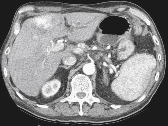
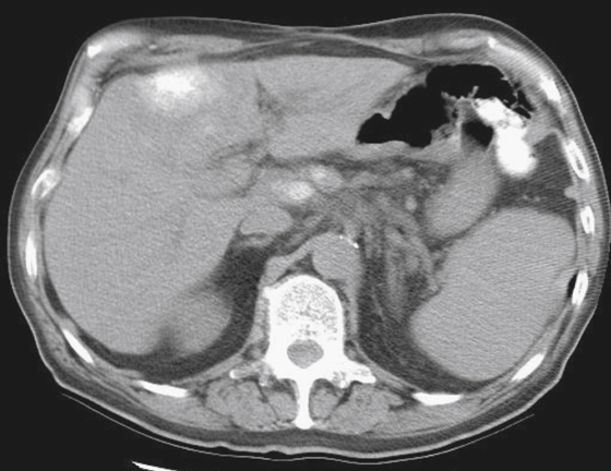
The CT image demonstrates calcified liver and lymph nodes lesions (see figures). Primary liver tumors may calcify and typically are solitary. Infectious processes that have involved the liver, such as granulomatous infections, also can produce liver calcifications. Parasitic diseases, such as echinococcal cysts, can cause calcification. However, the illustrated case shows multiple lesions that are either partially or completely calcified, which is strongly suggestive of metastatic disease.
Metastases to the liver rarely calcify. In the adult population, calcification of liver metastases is typically the result of a mucinous adenocarcinoma, which produces a psammomatous type of calcification that is detectable on CT scans. Most often, mucinous adenocarcinoma is found in the colon. Other sites of mucinous carcinoma include the pancreas, stomach, and ovaries. Tumors such as osteogenic sarcoma and chondrosarcoma can produce calcification or ossification, and their metastases could have this appearance as well. In children the most likely cause is neuroblastoma; up to 25% of neuroblastoma metastases calcify. Tumors outside the abdominal cavity rarely produce calcified liver metastases, but lung tumors, breast tumors, melanomas, and testicular tumors produce these lesions on rare occasion. Tumors that have been treated with chemotherapy or radiation also can calcify, although admittedly this presentation is rare. Calcification has been reported in various treated tumors and in treated lymphoma of the liver.


