CASE 103
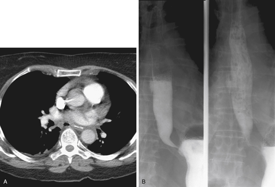
History: A 61-year-old woman developed chest pain after an episode of vomiting.
1. Which of the following should be included in the differential diagnosis of the imaging finding shown in Figure A? (Choose all that apply.)
E. Esophageal pseudodiverticulosis
2. This patient has developed a distal esophageal mucosal tear and intramural dissection after forceful retching. What is this syndrome?
3. Which of the following statements regarding Mallory-Weiss tears is true?
B. They are caused by vomiting or retching.
C. They are usually associated with alcohol abuse.
D. Left untreated, they usually progress to perforation.
4. Which of the following statements regarding Boerhaave’s syndrome is true?
A. It usually resolves spontaneously.
B. Endoscopy is the investigation of choice.
C. It is most commonly due to vomiting or straining.
D. The treatment of choice in all cases is surgical.
ANSWERS
CASE 103
Esophageal Tear
1. A, C, and D
2. B
3. C
4. C
References
de Lutio di Castelguidone E, Merola S, Pinto A, et al: Esophageal injuries: spectrum of multidetector row CT findings. Eur J Radiol. 2006;59:344–348.
Cross-Reference
Gastrointestinal Imaging: THE REQUISITES, 3rd ed, p 19.
Comment
Air in the esophageal wall is an uncommon finding. It is extremely difficult to see on plain film or even on barium esophagograms. However, the finding is easily identified on CT (see figures). The Mallory-Weiss patient can have air in the esophageal wall if an open mucosal tear exists (see figures). These patients usually are alcoholics (but not always). The condition has also been described in a few patients with severe hiccups. They present with blood-tinged vomiting and chest pain. The tears are almost always treated conservatively, and about 95% spontaneously heal. Patients with portal hypertension and a Mallory-Weiss tear are a more serious problem. A full-thickness tear of the distal esophagus (Boerhaave syndrome) is a surgical emergency and requires immediate intervention. The morbidity rate is high. Endoscopic instrumentation occasionally is the cause of mucosal tears. However, the risk of iatrogenic injury to the mucosa will probably increase as newer methods of endoscopic ablation of Barrett’s metaplasia are being tried with varying degrees of success and associated complications (including pneumatosis). Some of these procedures include photodynamic therapy, laser therapy, electrocoagulation, argon plasma coagulation therapy, and mucosal surface resection. In addition, radiofrequency and cryotherapy are being evaluated.


