CASE 102
History: A 16-year-old boy presents with abdominal discomfort, nausea, and weight loss.
1. Which of the following should be included in the differential diagnosis of the imaging finding shown in Figure A? (Choose all that apply.)
2. Which of the following statements regarding ascariasis is true?
A. The majority of sufferers come from Africa.
B. It is the most common intestinal parasitic infection.
C. The disease spreads most easily during the dry season.
D. The disease is transmitted through dogs as the animal reservoir.
3. What is the most common abdominal visceral complication of this infestation?
4. Which of the following is not a recognized manifestation of pulmonary involvement by this organism?
ANSWERS
CASE 102
Ascariasis of the Small Bowel
1. B, C, and D
2. B
3. B
4. C
References
Herlinger H, Ekberg TO. Other inflammatory conditions of the small bowel. In: Gore RM, Levine MS, eds. Textbook of Gastrointestinal Radiology. 2nd ed. Philadelphia: WB Saunders; 2000:746–758.
Cross-Reference
Gastrointestinal Imaging: THE REQUISITES, 3rd ed, p 135.
Comment
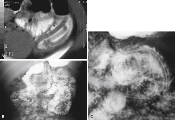
One of the most common parasitic infections is produced by the nematode Ascaris lumbricoides. It infects a major proportion of the world’s population. With the ease of worldwide travel, as well as immigration, this parasite is encountered in all areas of the world. The pathway of infection is quite complicated. The eggs of this parasite are ingested when infected water or food is consumed. In the gastrointestinal tract (small bowel) the larvae hatch and burrow through the intestinal wall. From there, they reach the portal venous system and travel to the liver and the lungs. The larvae then reach the bronchial system, where they can be found in the sputum, and reach the intestines by being swallowed in the sputum. Once they again reach the intestines, they grow into adult worms, which can be quite large.
The parasite produces diseases in many ways. The larvae may produce a local hypersensitivity reaction, which is particularly evident when they are in the lungs. When they are in the intestines, the worms cause nutritional deficiencies. As the worms grow, the large mass of the worms can produce obstruction and even appendicitis. Perforation of the intestines with peritonitis can even occur. Often the patient’s symptoms are quite vague, with occasional pain and diarrhea. The worms can also migrate into the biliary system, where they can produce cholangitis or pancreatitis because of their size as well as a local inflammatory response.
Radiologically the worms are visible on barium studies because they are so large (see figures). There may be a single worm, or they can occur in large masses. A hallmark of these worms is that they ingest the barium during the examination, and then barium outlines the intestinal tract of the worms, as is evident in this case (see figures).


