CASE 102
1. What etiologies can cause this finding? (Choose all that apply.)
A. Coronary artery bypass graft surgery
B. Infection
D. Uremia
2. What is the pericardial abnormality?
A. Effusion
B. Thickening
D. Nodularity
3. Given the symptoms, what is the most likely diagnosis?
C. Tamponade
D. Tumor
4. What is the most appropriate treatment?
A. Antibiotics
ANSWERS
References
Wang ZJ, Reddy GP, Gotway MB, et al. CT and MR imaging of pericardial disease. Radiographics. 2003;23(Spec No):S167–S180.
Yared K, Baggish AL, Picard MH, et al. Multimodality imaging of pericardial diseases. JACC Cardiovasc Imaging. 2010;3(6):650–660.
Cross-Reference
Cardiac Imaging: The REQUISITES, ed 3, pp 269–271.
Comment
Etiology and Physiology
Constrictive pericarditis occurs when limitation in diastolic ventricular filling leads to equalization of atrial and ventricular pressures, which is known as constrictive/restrictive physiology. Patients have symptoms similar to congestive heart failure. Physical examination may demonstrate the classic Kussmaul sign, which is the paradoxical elevation of jugular venous pressure on inspiration. Causes of constrictive pericarditis include open heart surgery, radiation therapy, uremic pericarditis, viral pericarditis, and tuberculous pericarditis (less common in industrialized countries).
Treatment
Constrictive pericarditis and restrictive cardiomyopathy have similar clinical presentations and findings on echocardiography and cardiac catheterization. However, it is important to differentiate between these two diseases because patients with constrictive disease usually benefit from pericardiectomy, whereas restrictive cardiomyopathy has a poor prognosis and must be managed medically or with heart transplantation.
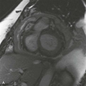
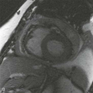
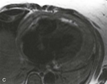
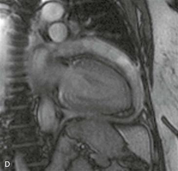
Imaging
In the clinical setting of constrictive/restrictive physiology, pericardial thickening (≥4 mm) as demonstrated on MRI can establish the diagnosis of constrictive pericarditis (Figs. A–D). Diastolic septal dysfunction (septal bounce) on cine MRI is another key finding. Ancillary findings may be present, including dilation of the inferior vena cava, hepatic veins, and right atrium, with a narrow, tubular right ventricle. Pericardial thickening can occur in the absence of constrictive physiology. The diagnosis of constrictive pericarditis can be established only in the appropriate clinical setting of constrictive/restrictive physiology.


