Published on 02/03/2015 by admin
Filed under Internal Medicine
Last modified 22/04/2025
This article have been viewed 953 times
Case 1
A 68-year-old man with a history of hypertension presented to the emergency department for evaluation of new-onset weakness and diaphoresis. He denied chest pain. He had no other coronary disease risk factors.
Laboratory examination was normal except for leukocytosis (WBC = 22,000). He left against medical advice but returned 2 days later for persistent symptoms. Exercise SPECT MPI was requested.
The baseline ECG shows normal sinus rhythm with slightly increased QRS voltage consistent with left ventricular hypertrophy.
He exercised on a Bruce protocol for 6:32 minutes, achieving an estimated workload of 7.7 METs. The peak exercise heart rate was 146 beats/min (96% of maximum age-predicted heart rate). No chest pain was reported, and the blood pressure response to exercise was normal.
The peak exercise ECG demonstrated sinus tachycardia with 4 mm of downsloping ST-segment depression suggestive of ischemia.
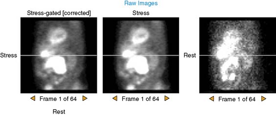
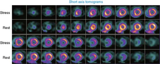
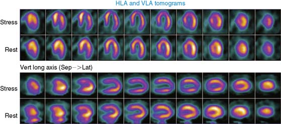
The postexercise (above) and resting (below) raw images are shown. Image quality appears adequate.
The poststress images demonstrate a mild defect in the anterior and anterolateral regions and a moderately severe defect in the apical region. Resting images demonstrate nearly complete defect reversibility.
On comparison of the patient’s perfusion pattern to a male-specific normal database, the apical perfusion is deemed to be abnormal (the apical defect involves 8% of the left ventricular myocardium). On comparison to resting images, significant defect reversibility is evident (7% of the left ventricular myocardium).
Postexercise gated SPECT images demonstrated low-normal global left ventricular systolic function (LVEF = 50%) with borderline increased left ventricular end-diastolic (normal < 120 mL) and end-systolic (normal < 70 mL) volumes.
The SPECT MPI results were reviewed in the clinical context of the patient’s presenting symptoms. The presentation was atypical for an acute coronary syndrome and more consistent with an infectious etiology. The SPECT MPI results suggested the presence of inducible ischemia, though the perfusion abnormality appeared to be confined to a small portion (<10%) of the left ventricle. Medical therapy for CAD was therefore contemplated as a reasonable approach. However, the exercise ECG parameters suggested that the patient may be at high risk for cardiac events. The computed Duke treadmill score (DTS) was −13 (high-risk category). The DTS was calculated as: (7 min exercise) − (5 × 4 mm ST depression) − (4 × 0 anginal index*) = −13. Because of the high-risk DTS, he was referred for coronary angiography.
The coronary angiogram revealed 80% stenosis of the distal left main artery (LM), with mild (10%) diffuse stenoses in the LAD, LCX, and RCA. Coronary artery bypass grafting was recommended.
This case illustrates the detection of isolated LM stenosis by SPECT MPI. The sensitivity of contemporary SPECT MPI for detecting isolated LM stenosis is low (<50%). Frequently, LM disease is detected “fortuitously” because there is a second focal stenosis present in a remote coronary artery that produces a perfusion defect and leads to diagnostic coronary angiography. Patients with isolated LM stenosis (who lack an additional flow-limiting stenosis elsewhere) frequently have minimal to no perfusion defect detected on SPECT MPI.
Several imaging (and nonimaging) markers have been proposed to enhance detection of LM stenosis with SPECT MPI. These markers include:
In this patient, clues to the presence of left main stenosis included marked ST depression and an increase in stress/rest LV cavity ratio (1.49; normal < 1.22). In the future, it is hoped that routine measurement of absolute (rather than relative) myocardial blood flow with rubidium-82 PET MPI may improve our ability to detect isolated LM stenosis (and other conditions in which “balanced” myocardial hypoperfusion occurs during stress).
Clinical Nuclear Cardiology State of the Art and Future Directio
WhatsApp us