CASE 1
History: No patient history is available.
1. What should be included in the differential diagnosis for ground-glass opacity? (Choose all that apply.)
B. Pneumonia
D. Adenocarcinoma in situ (bronchioloalveolar cell carcinoma)
2. If this patient presented with fever and a productive cough, what is the most likely diagnosis?
A. Right upper lobe pulmonary edema
D. Adenocarcinoma in situ (bronchioloalveolar cell carcinoma)
3. If this patient presented with severe chest pain and no infectious symptoms, what would be the most likely diagnosis?
A. Right upper lobe pulmonary edema
D. Adenocarcinoma in situ (bronchioloalveolar cell carcinoma)
4. If acute mitral regurgitation is the suspected diagnosis based on the chest radiograph and clinical presentation, what is the next best step in management?
A. CT
D. MRI
ANSWERS
Reference
Schnyder PA, Sarraj AM, Duvoisin BE, et al. Pulmonary edema associated with mitral regurgitation: prevalence of predominant involvement of the right upper lobe. AJR Am J Roentgenol. 1993;161(1):33–36.
Cross-Reference
Cardiac Imaging: The REQUISITES, ed 3, pp 191–194.
Comment
Pathophysiology
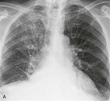
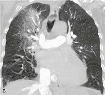
Asymmetric right upper lobe pulmonary edema is seen in 9% of adults and 22% of children with severe mitral regurgitation. In adults, it is usually caused by a flail posterior valve leaflet secondary to myocardial infarction. A flail posterior leaflet causes the mitral regurgitant jet to be preferentially directed into the right superior pulmonary vein; this leads to focal increased hydrostatic pressure and pulmonary edema within the right upper lobe. In the setting of chest pain, acute mitral regurgitation must be considered and can be confirmed with echocardiography.
Imaging
The radiograph (Fig. A) and CT scan (Fig. B) show asymmetric right upper lobe ground-glass opacity. The differential diagnosis varies depending on the patient’s clinical presentation. In the setting of fever and productive cough, the imaging findings are consistent with atypical pneumonia. In this case, the patient had severe chest pain and acute myocardial infarction. Echocardiography confirmed severe mitral regurgitation.


