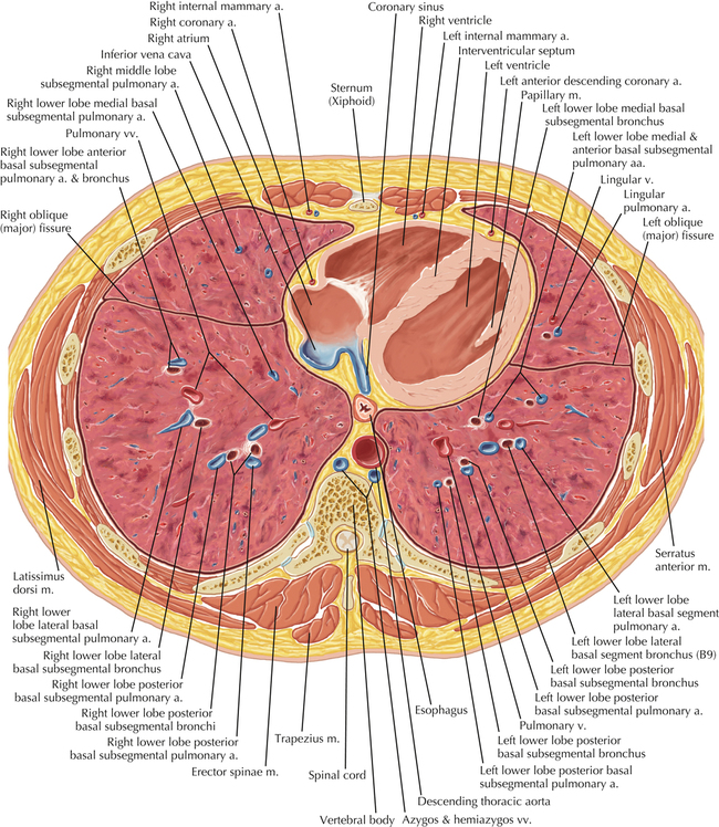
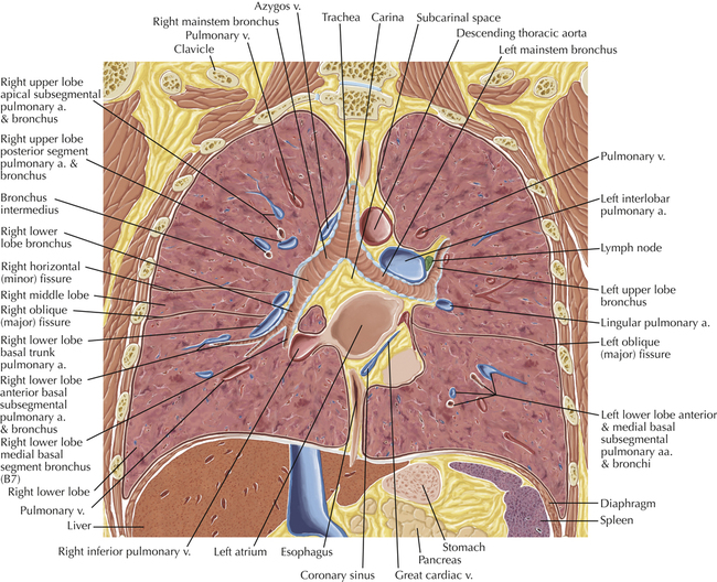
Thoracic Soft Tissue and Lung
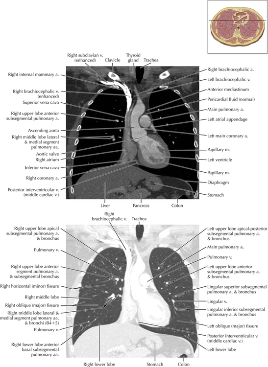
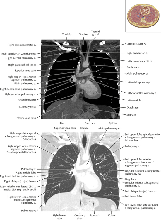
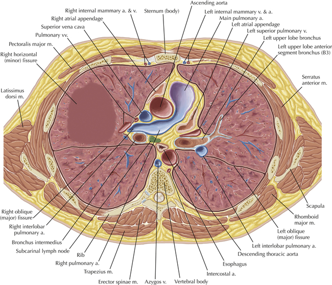
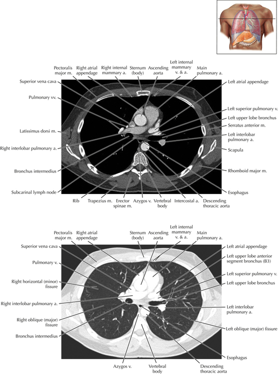
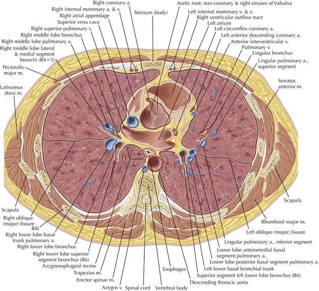
Soft Tissue and Lung Axial 3
Diagnostic Consideration
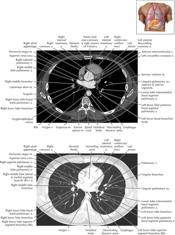
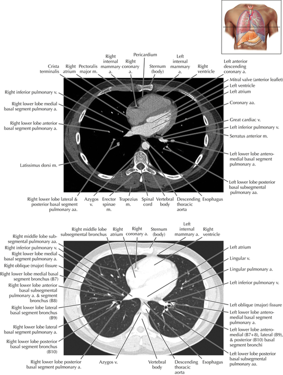
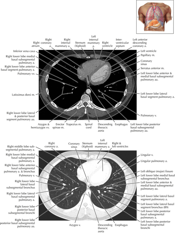
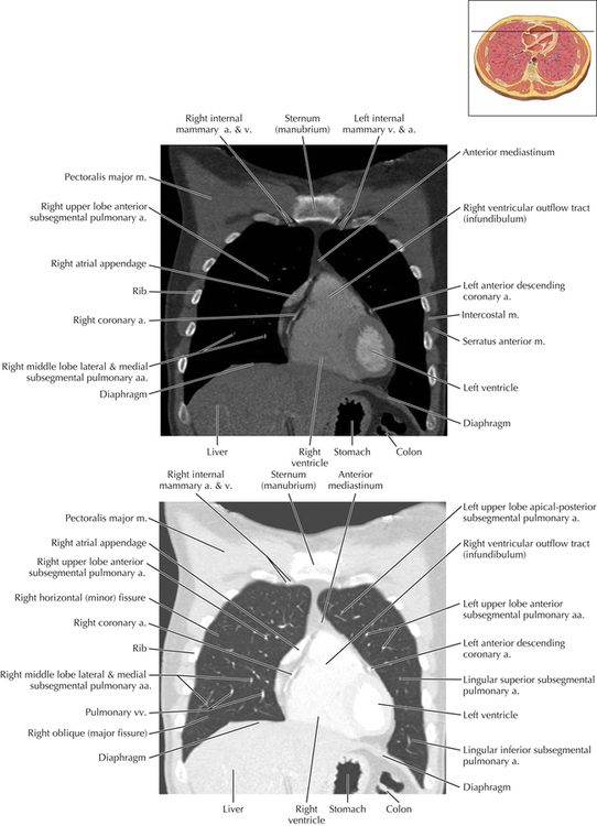
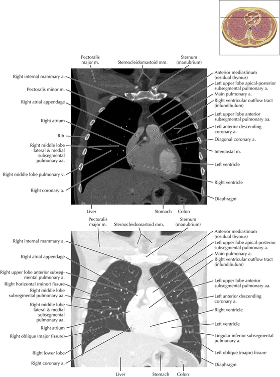
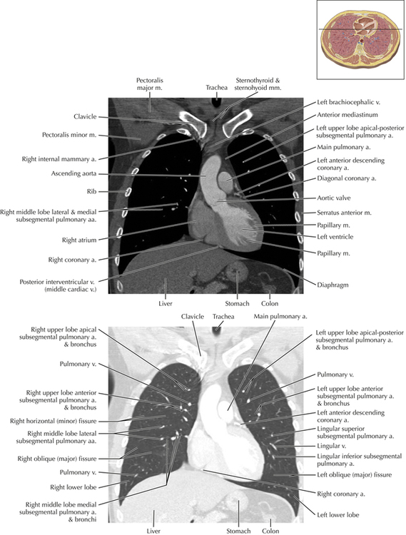
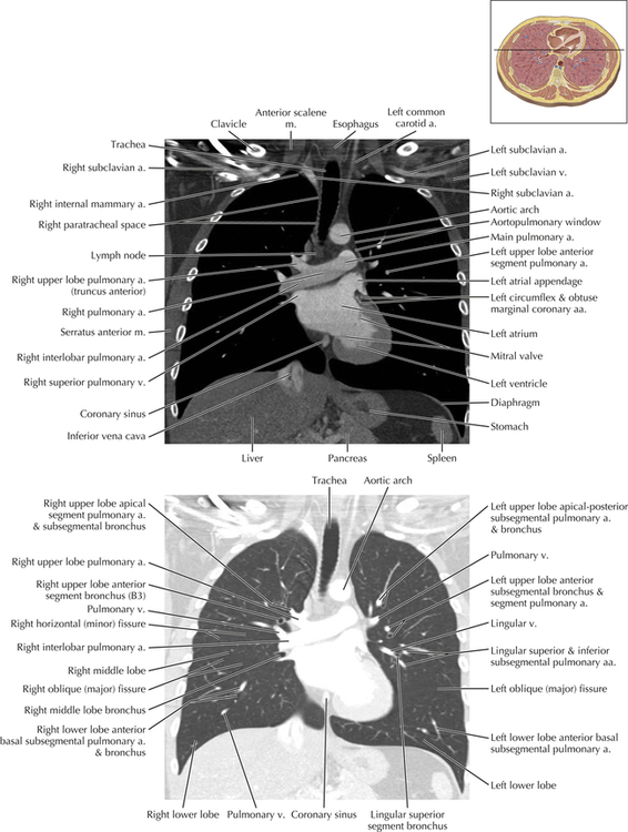
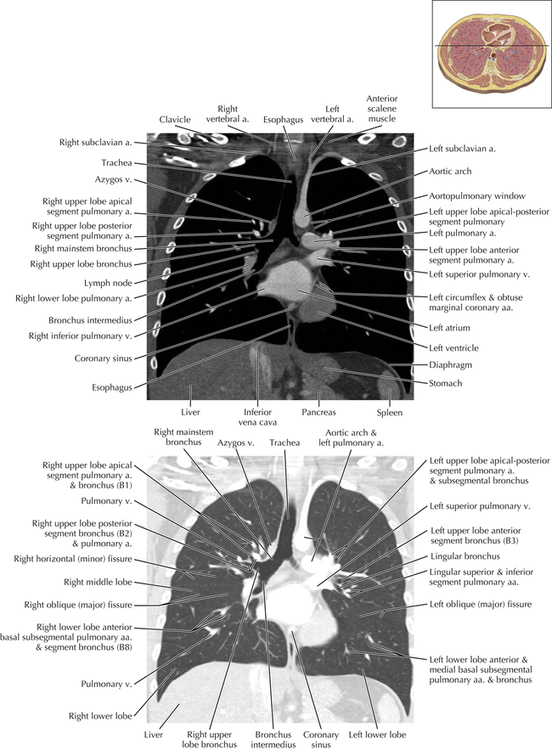
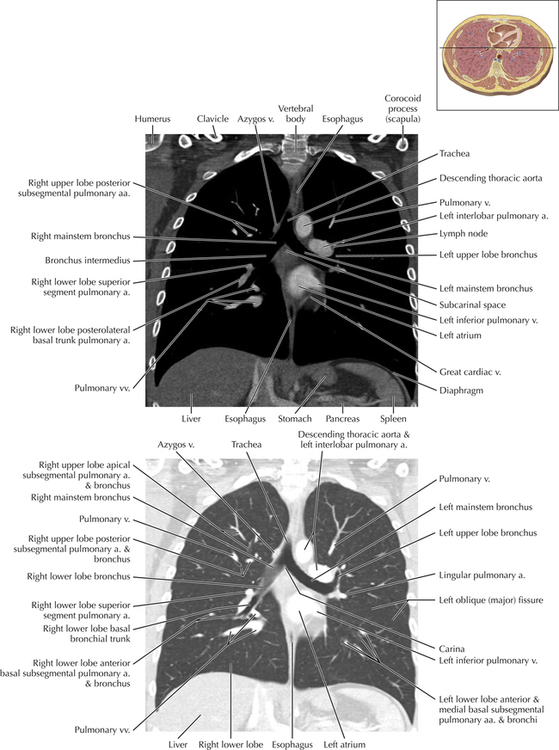
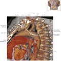
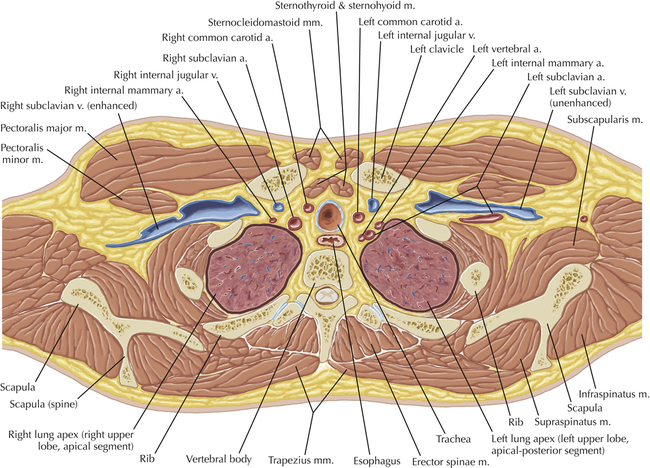
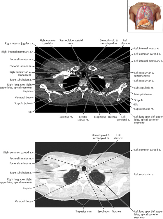
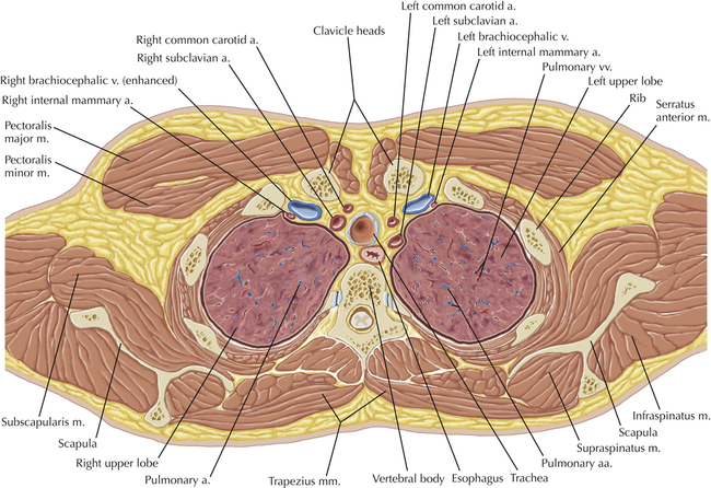
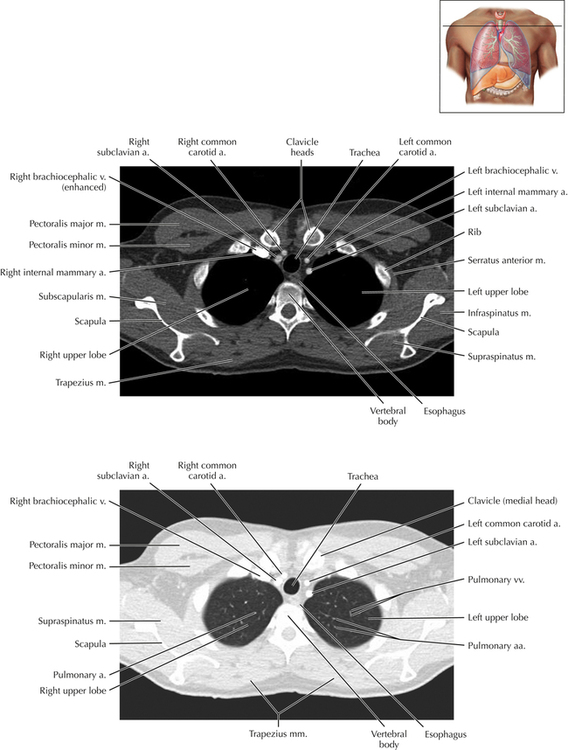
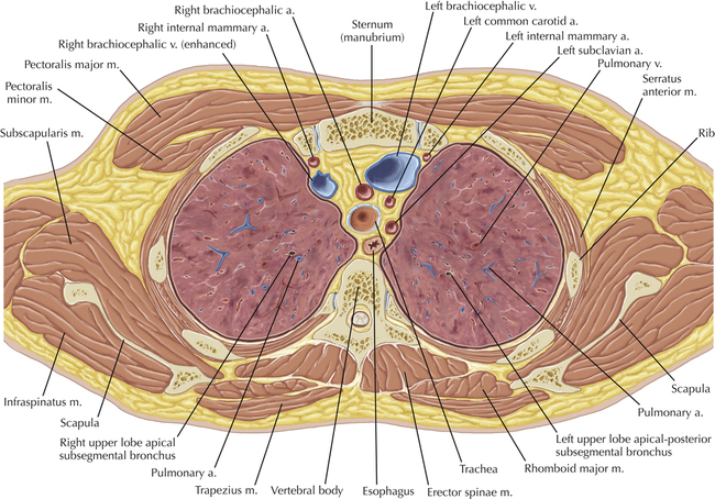
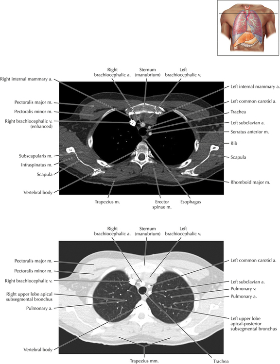
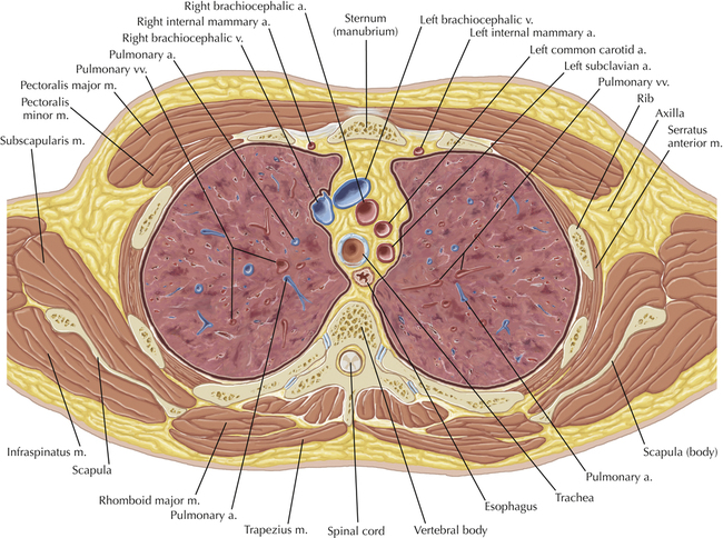
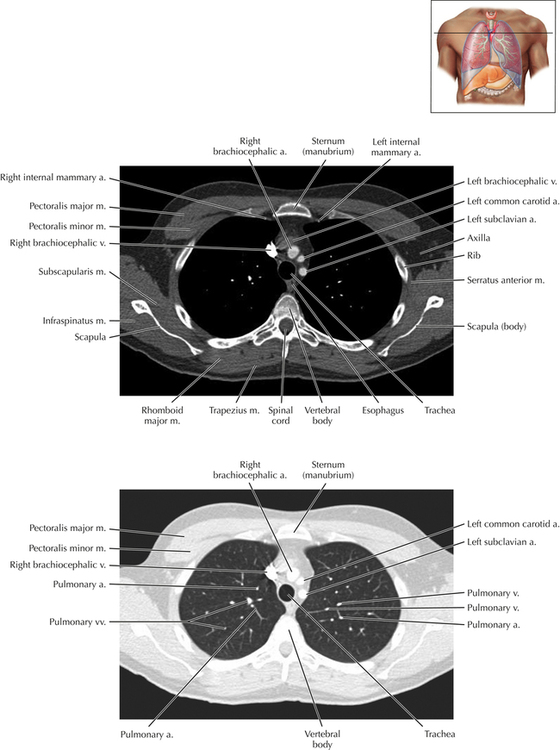
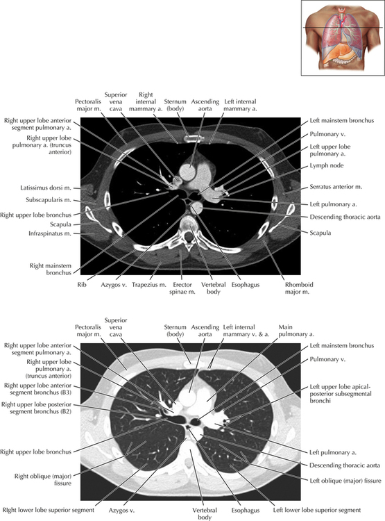
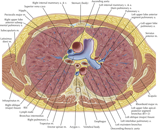
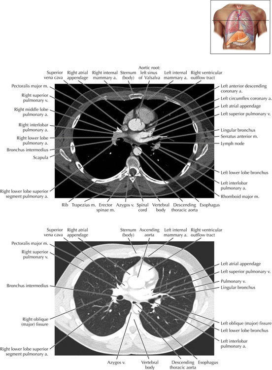
Thoracic CT scans may be performed with or without the intravenous injection of iodinated contrast. Contrast-enhanced thoracic CT scanning is usually performed following injection into an upper extremity vein, with imaging started at 30 to 40 seconds after the injection is begun, depending on the specific application. In this patient, contrast was injected via the right upper extremity, resulting in dense enhancement of the right subclavian vein (and the right brachiocephalic vein on Axial 5 and subsequent images); the left subclavian vein (and left brachiocephalic vein on Axial 5 and subsequent images) remains unenhanced in this patient because imaging was begun before injected contrast had sufficient time to circulate throughout the body and reach the left subclavian vein.
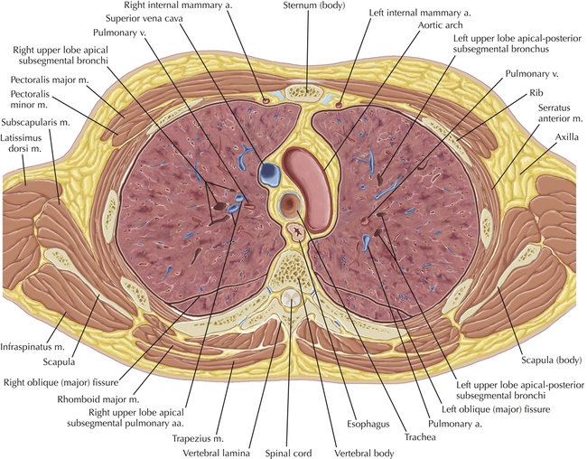
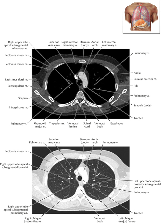
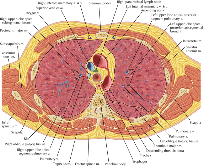
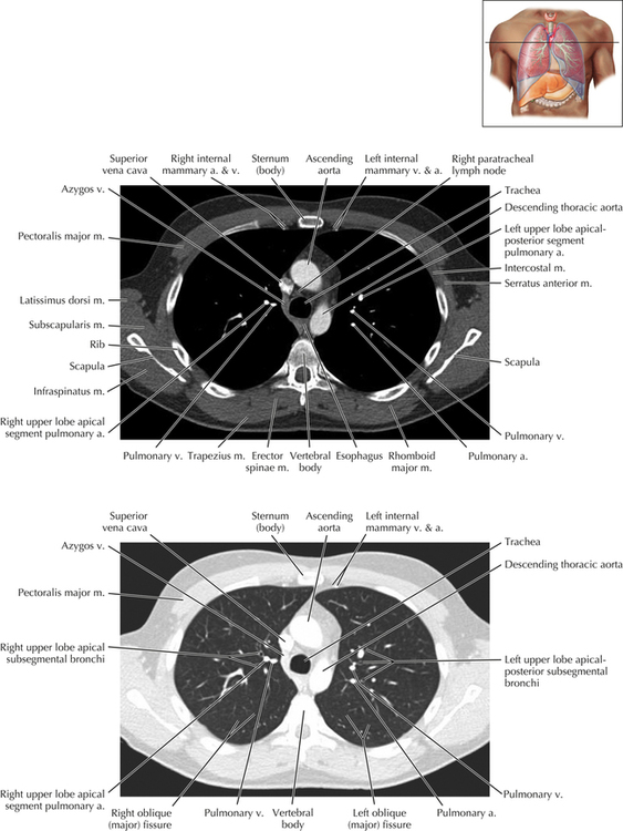
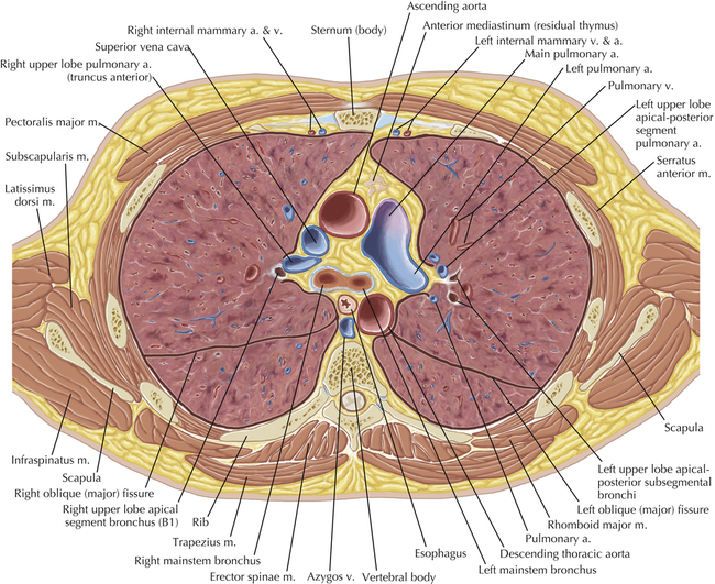
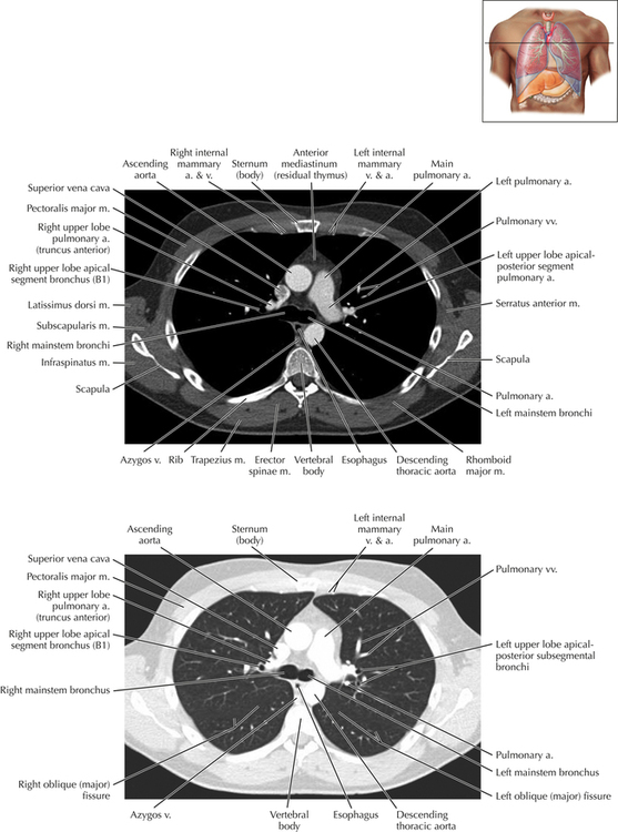
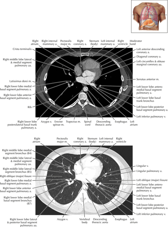
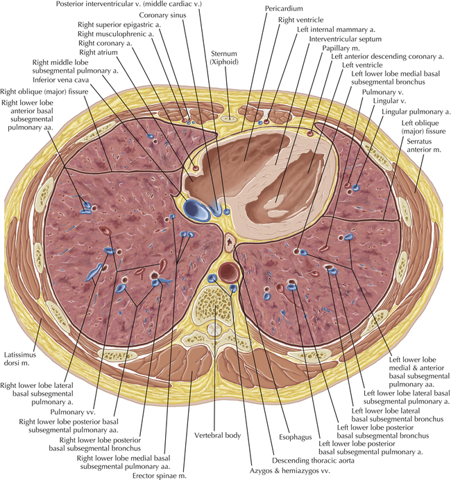
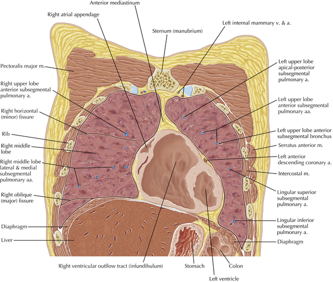
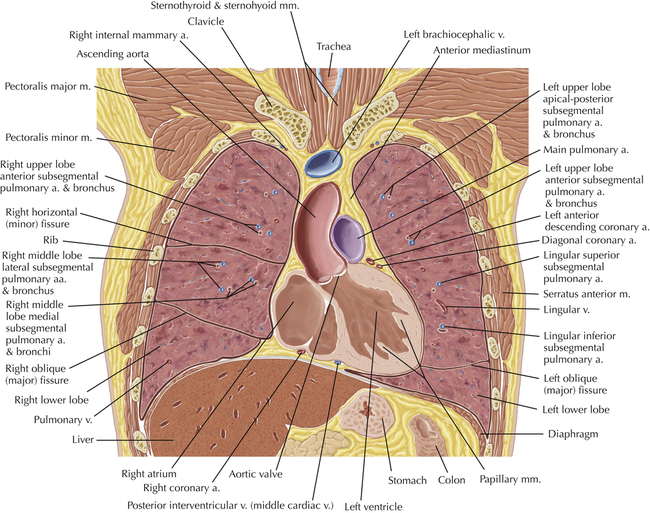
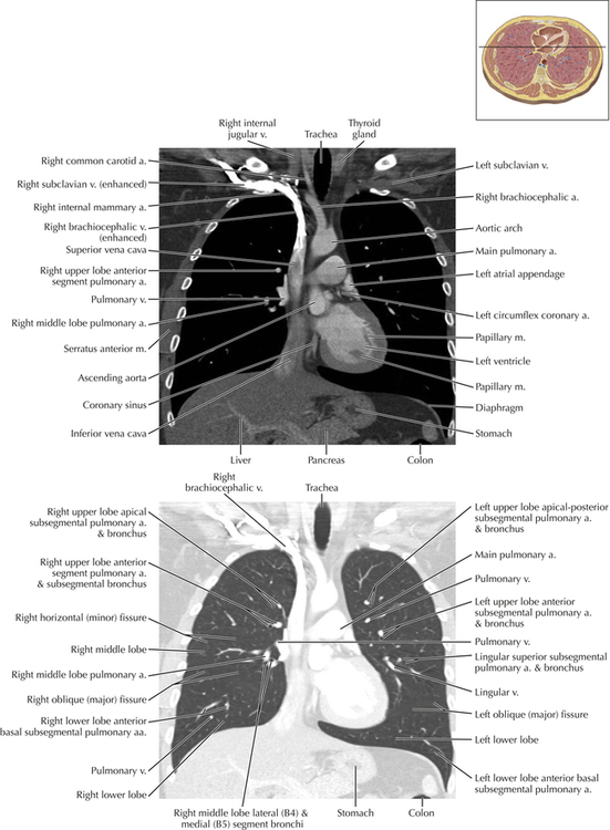
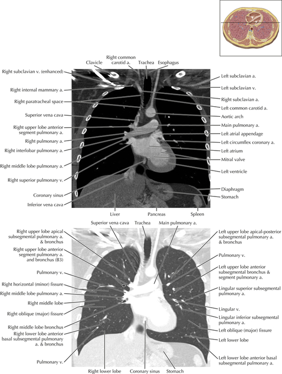
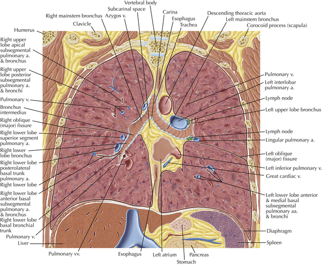
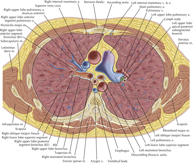
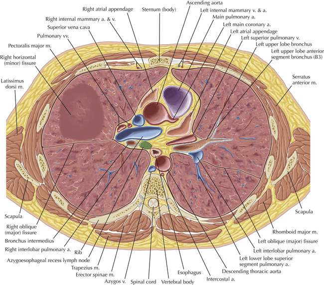
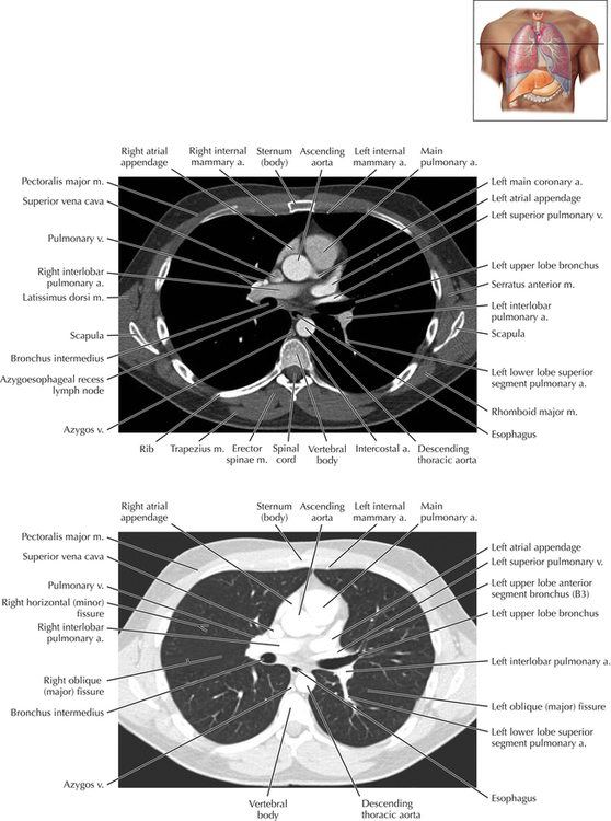
Soft Tissue and Lung Axial 16
Normal Anatomy
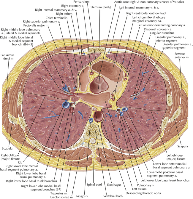
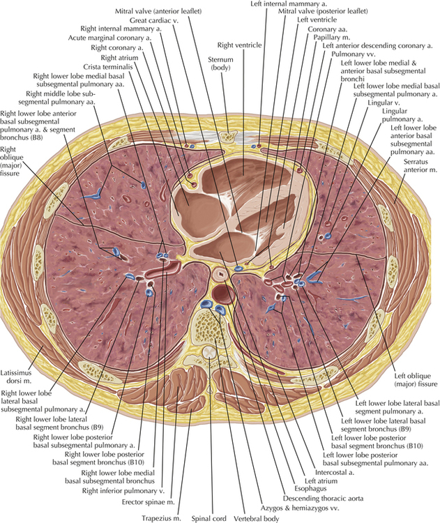
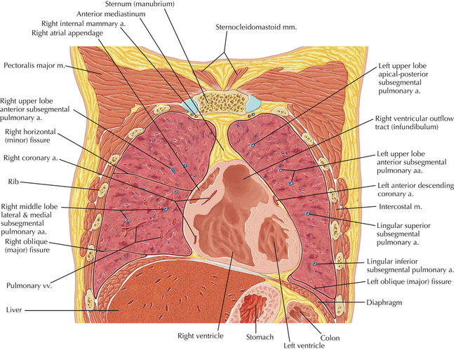
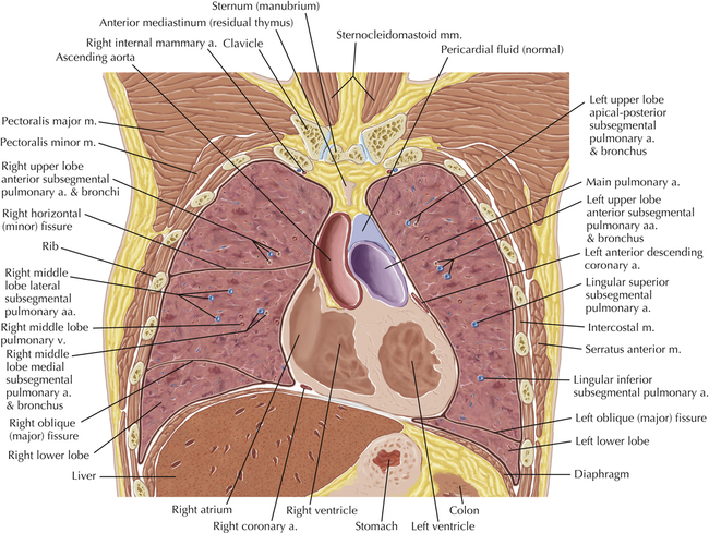
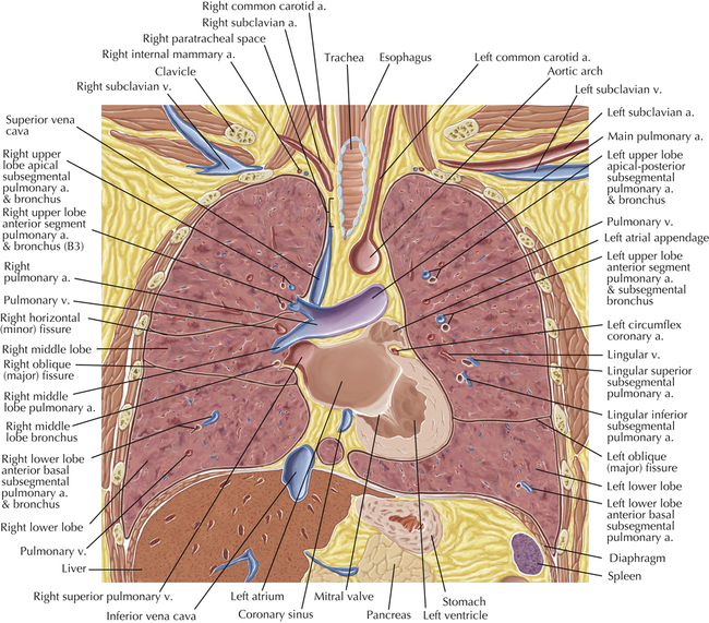
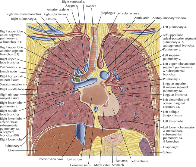
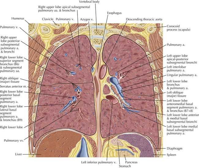
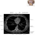
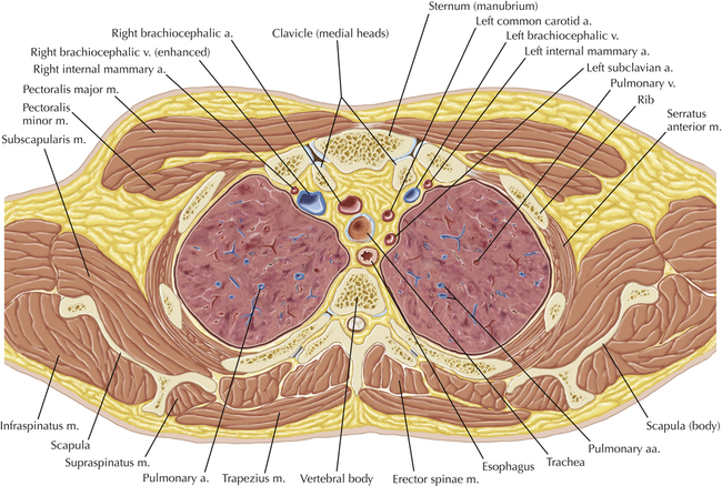
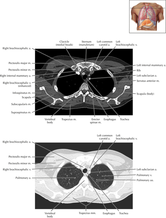
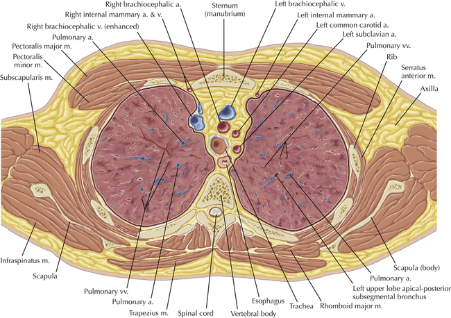
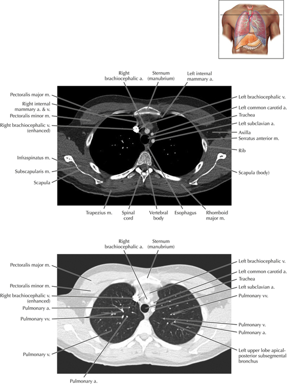
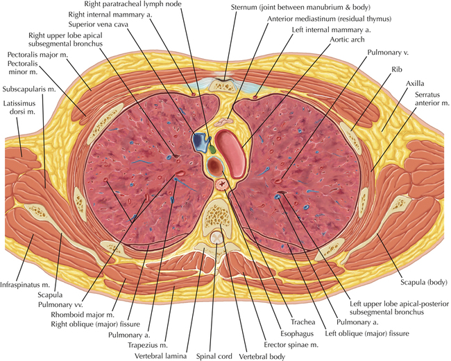
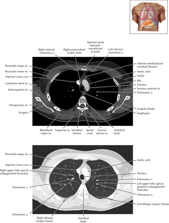
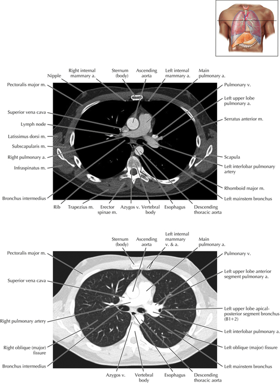
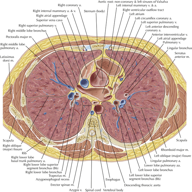
At this level, the segmental bronchi within the right upper lobe are now visible. Segmental bronchi are commonly named for the anatomic segment they supply (e.g., apical segment right upper lobe bronchus). Another nomenclature system for segmental bronchi may occasionally be encountered, as described by the Federative Committee on Anatomical Terminology in Terminologia Anatomica. This terminology identifies the segmental bronchi according to numbers (e.g., B1 = apical segment right upper lobe bronchus, B1+2 = apical-posterior segment left upper lobe bronchus; see Axial 19). Both nomenclature systems are illustrated in this text.
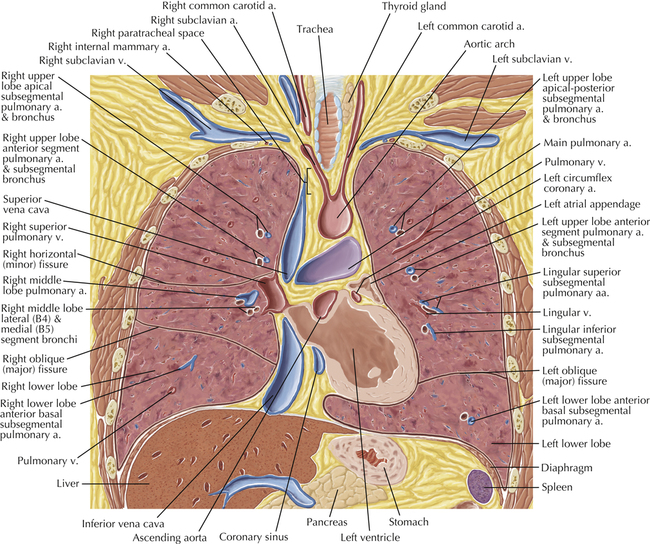
Normal Variant
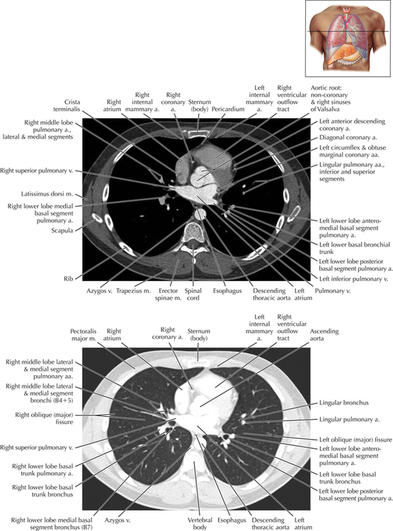
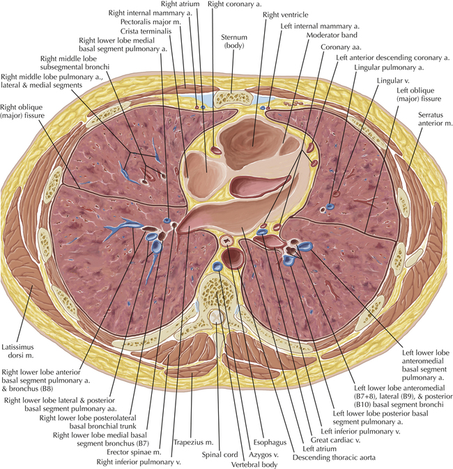
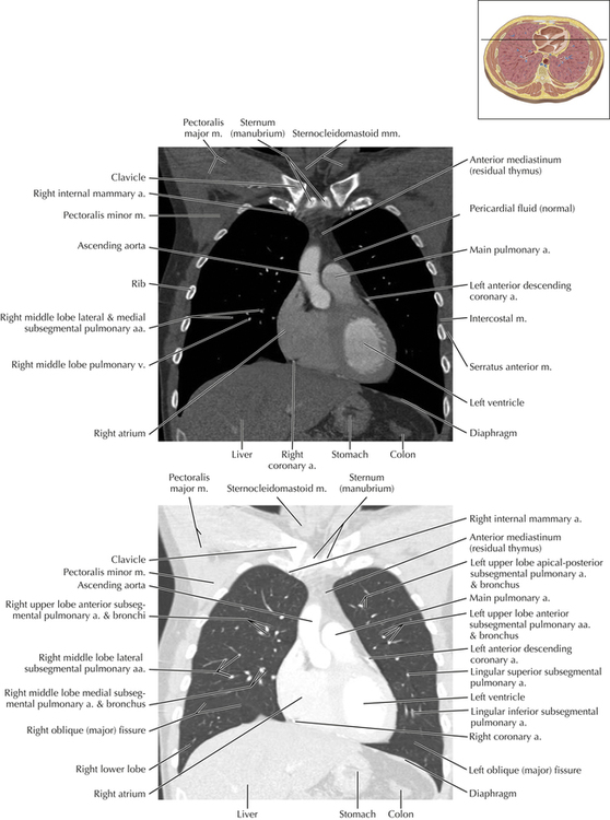
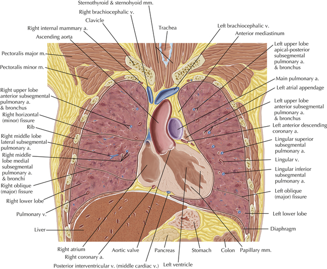
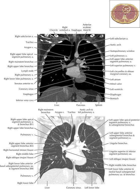
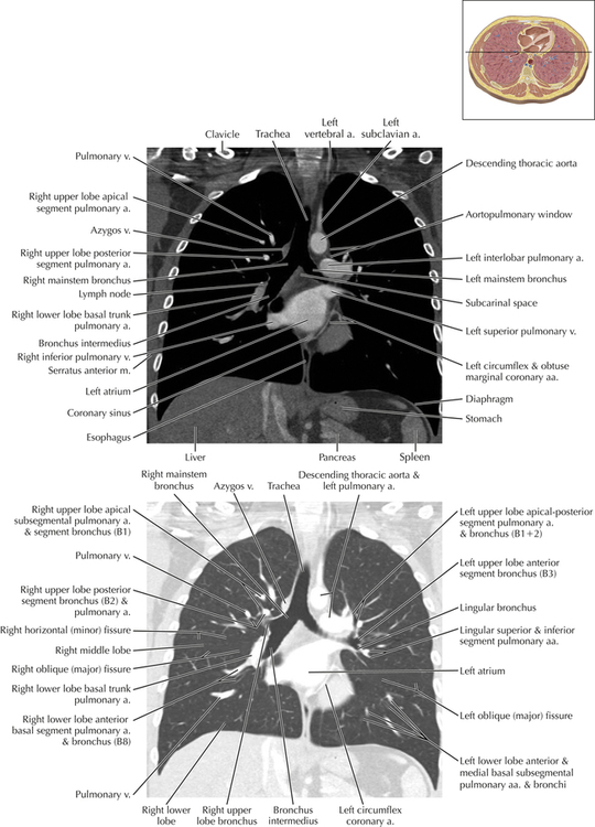
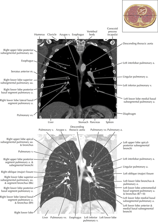
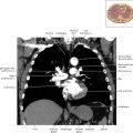
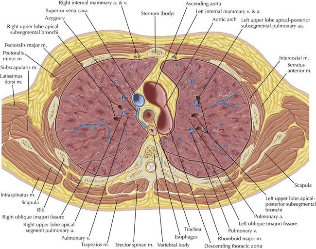
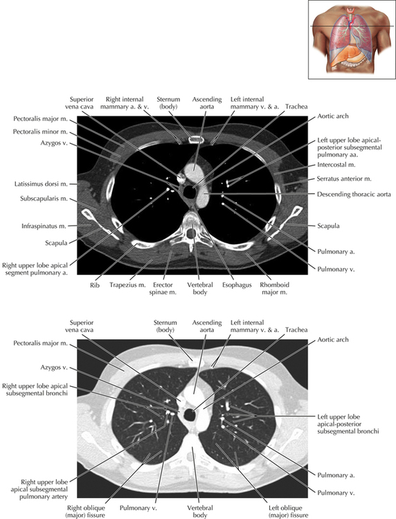
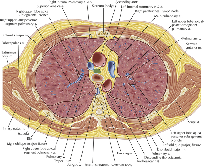
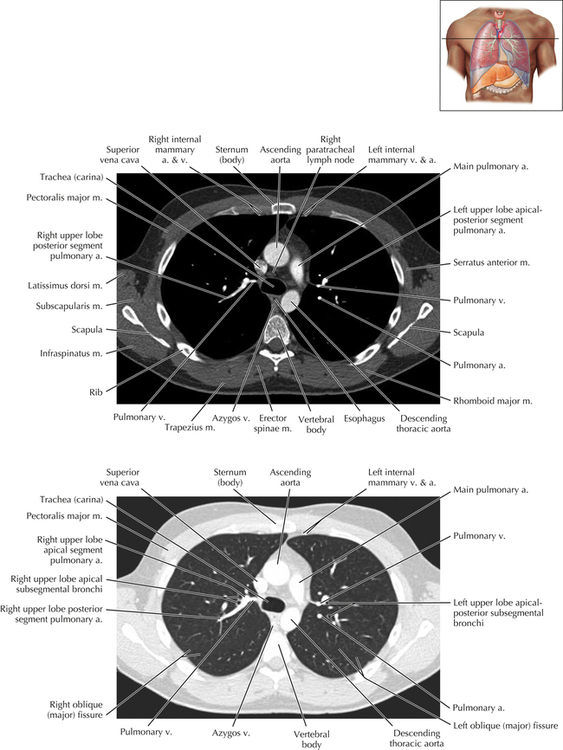
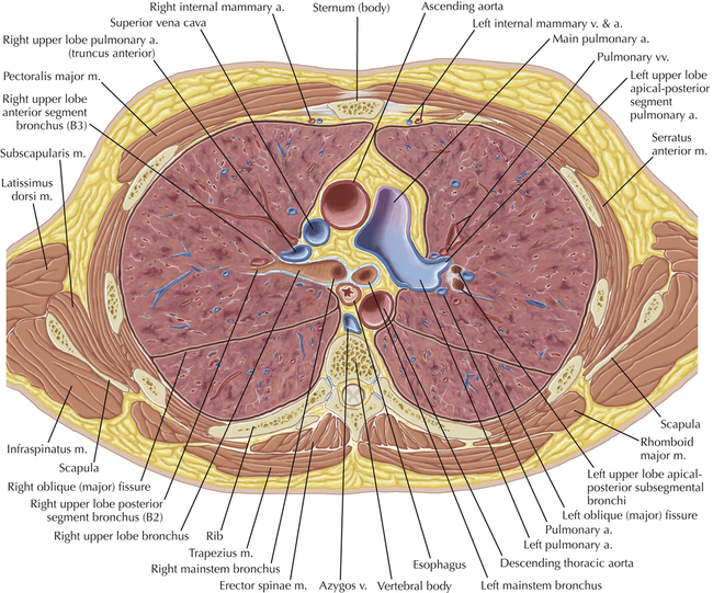
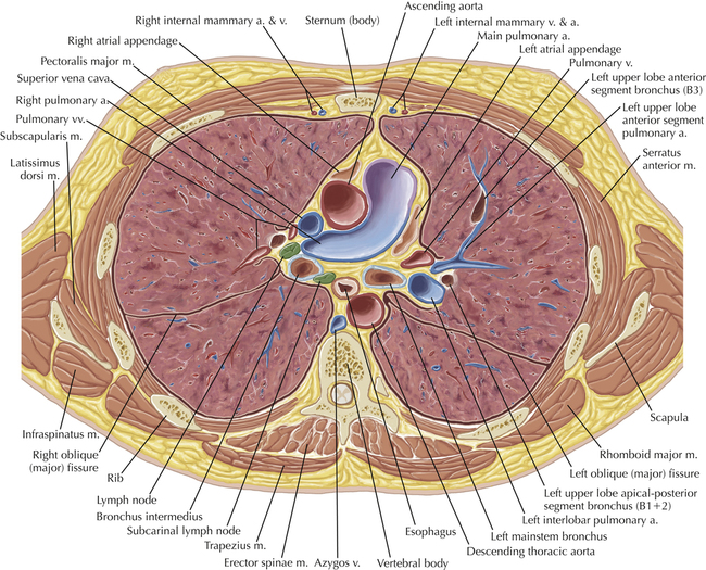
A number of variations in the branching pattern of the pulmonary arteries are recognized. In particular, variations in the branching pattern of the left upper lobe pulmonary artery are commonly encountered in clinical practice. In the most common pattern, the left upper lobe pulmonary artery gives rise to apical-posterior and anterior segmental branches in a manner analogous to the bronchial branching pattern. Less commonly, as in this patient, a posterior left upper lobe subsegmental artery arises directly from the left pulmonary artery. The left upper lobe pulmonary artery can be seen originating from the left pulmonary artery on Axials 17 and 18.
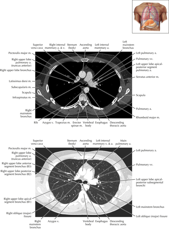
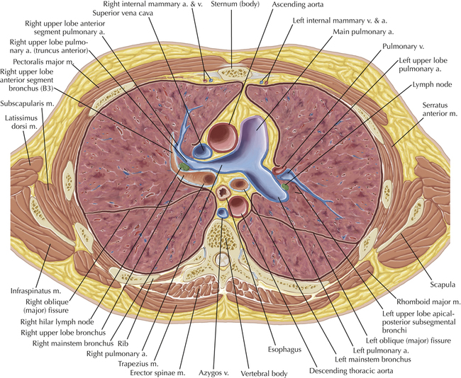
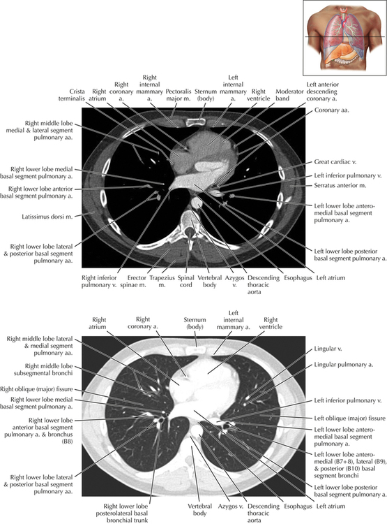
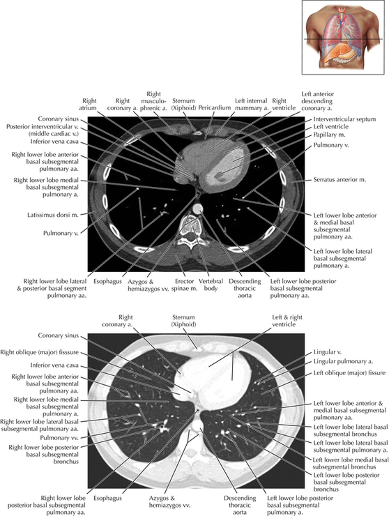
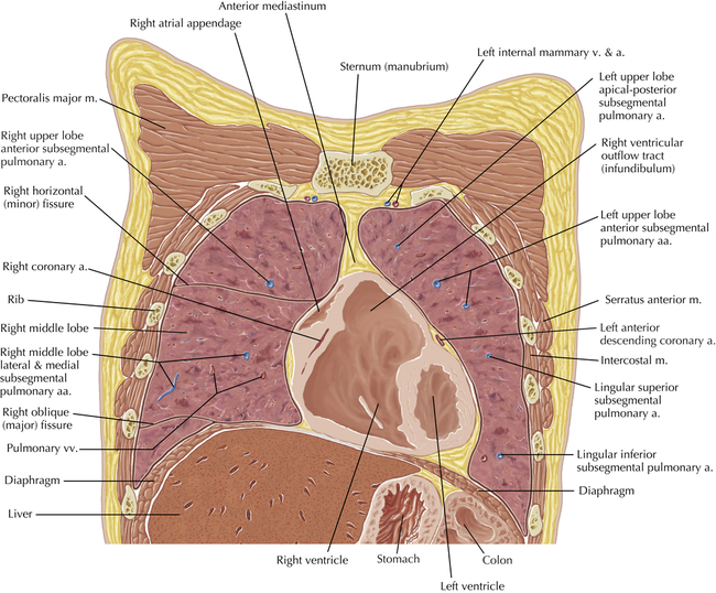
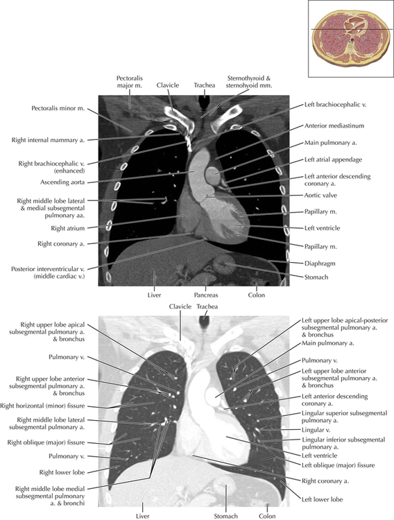
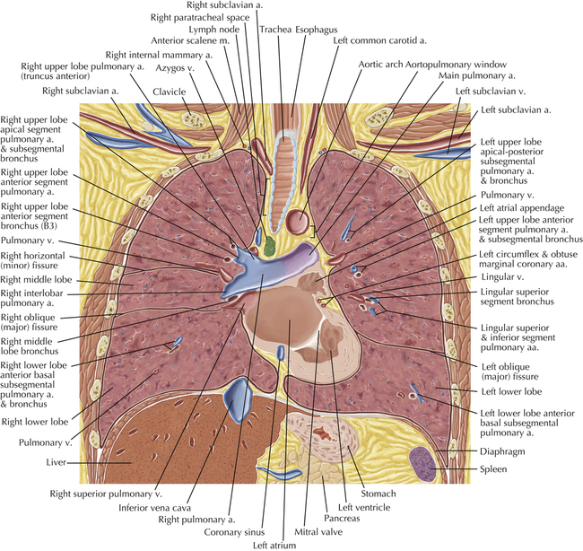
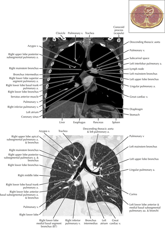
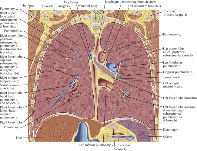
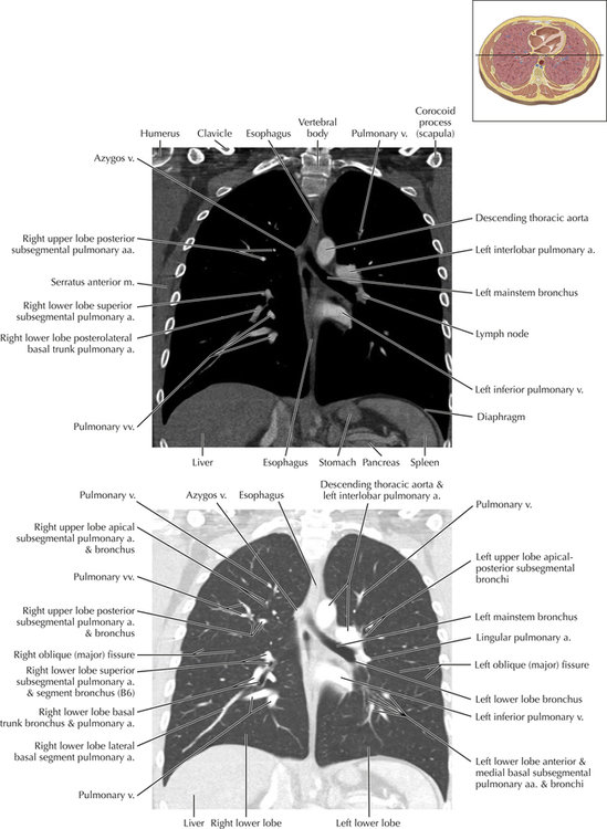
Soft Tissue and Lung Axial 25
Normal Variants
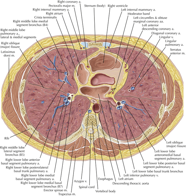
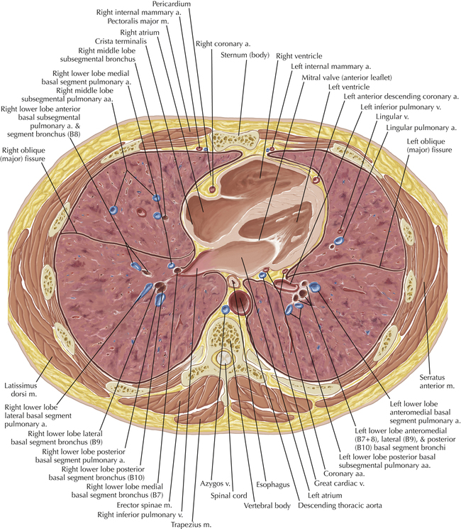
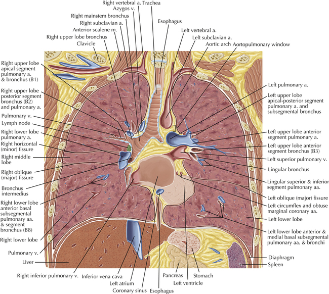
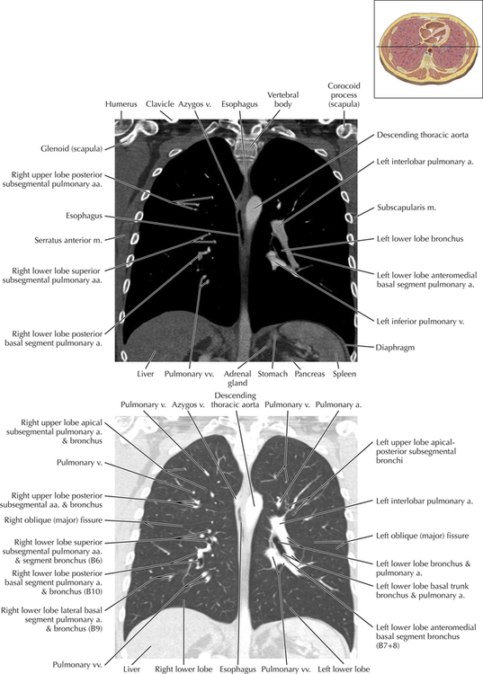
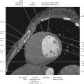
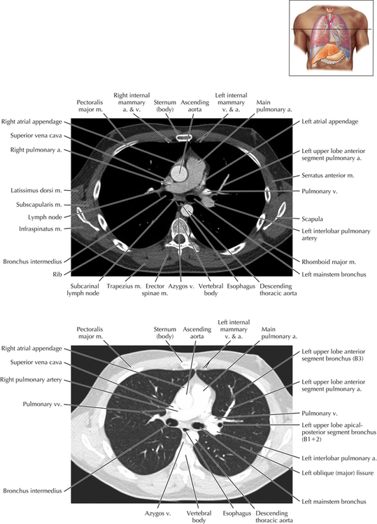
This level marks the origin of the left lower lobe segmental pulmonary arteries. Typically the left basal arterial trunk, inferior to the superior segmental left lower lobe pulmonary artery, branches into the anteromedial basal, lateral basal, and posterior basal segmental pulmonary arteries. In this patient, two, instead of three, basal segmental left lower lobe pulmonary arteries are visible. These vessels can be sequentially followed through Axial 29. On Axial 30, the lateral basal segmental lower lobe pulmonary artery is seen originating from the anteromedial basal segmental vessel. Although this pattern of segmental pulmonary arterial branching is uncommon, variations in the branching patterns of the pulmonary arteries are frequently encountered and usually of no clinical importance.