Rigler triad of small bowel (SB) obstruction, gas in biliary tree, and ectopic gallstone
• Location: Points of luminal narrowing of bowel; duodenum, ligament of Treitz, ileocecal valve, sigmoid colon
• CT findings
Gallstone surrounded by gas in bowel loop
– Cholesterol stones near-water density, often with calcified rim
Collapsed gallbladder, pneumobilia (gas within GB ± bile duct lumen)
Dilated bowel with transition to collapsed bowel at impacted gallstone
TOP DIFFERENTIAL DIAGNOSES
• Intussusception
• Dropped gallstone (intraperitoneal, not intraluminal)
PATHOLOGY
• Occurs with chronic cholecystitis
• Delayed (up to 2 months) complication of ERCP
• Diagnosis frequently delayed or missed
CLINICAL ISSUES
• Most common signs/symptoms: Intermittent acute colicky abdominal pain (20-30%), nausea, vomiting, fever, distension, obstipation
• Gallstone erodes inflamed GB wall, passes into GI tract (usually duodenum) → bowel obstruction
DIAGNOSTIC CHECKLIST
• Consider in elderly woman with recurrent RUQ pain, recently more severe and prolonged vomiting
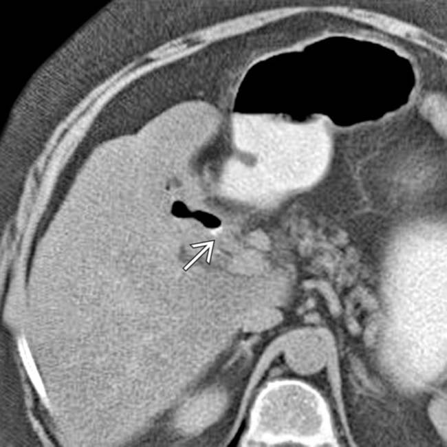
(Left) Axial CECT in a 65-year-old woman presenting with crampy abdominal pain demonstrates a thick-walled gallbladder with air within its lumen and a gas-filled fistula to the duodenum .
(Right) Axial CECT in the same patient identifies the obstructing gallstone , which is impacted in the distal jejunum. The proximal small bowel is dilated, while the distal SB and colon are collapsed.
(Left) Axial CECT in a patient presenting with bloating and abdominal pain demonstrates gas in the gallbladder , a typical finding in gallstone ileus, also known as gallstone obstruction.
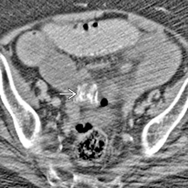
(Right) Axial CECT in the same patient demonstrates dilation of the proximal small bowel and decompressed, nondilated distal small bowel . The high-density obstructing gallstone is seen, as a laminated calcification, at the site of transition from the dilated to the decompressed bowel.
Axial NECT shows gas in the gallbladder adjacent to a large gallstone .
Axial NECT at a more caudal level in the same patient demonstrates a calcified gallstone obstructing the distal small bowel.
Anteroposterior radiograph shows dilated small bowel confirming obstruction, as well as gas in the gallbladder .
Axial CECT shows a dilated proximal small bowel and laminated filling defect at the transition point, which proved to represent an obstructing gallstone.
Axial CECT shows a cholecystoduodenal fistula from a prior episode of GSI. Note the gas in the gallbladder and the close proximity of the GB to the duodenum .
Axial CECT shows gas in a collapsed gallbladder. Note the fistula to the duodenum .
Axial CECT shows a large gallstone within the dilated small bowel. Note that the bowel distal to the gallstone is collapsed.
Axial CECT shows gas in a thick-walled collapsed gallbladder. Note the fistula to the duodenum.
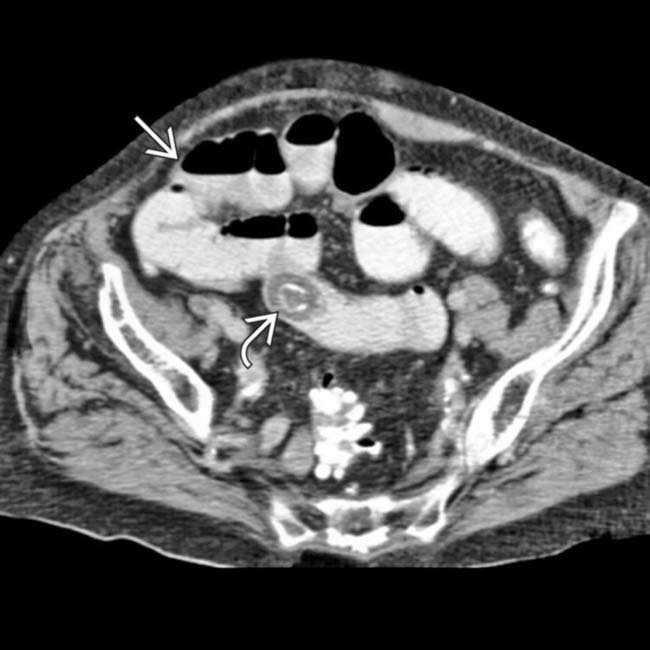
Axial CECT shows small bowel obstruction with a large, laminated gallstone “stuck” near the ileocecal valve.


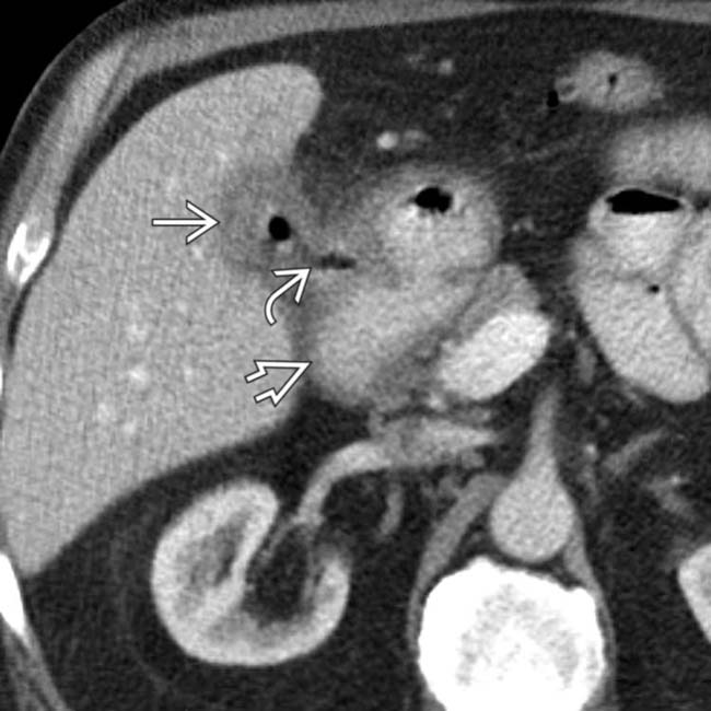
 with air within its lumen and a gas-filled fistula
  to the duodenum
  .
.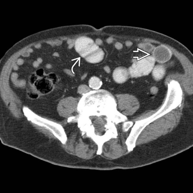
 , which is impacted in the distal jejunum. The proximal small bowel
  is dilated, while the distal SB and colon are collapsed.
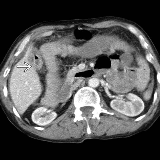
 , a typical finding in gallstone ileus, also known as gallstone obstruction.
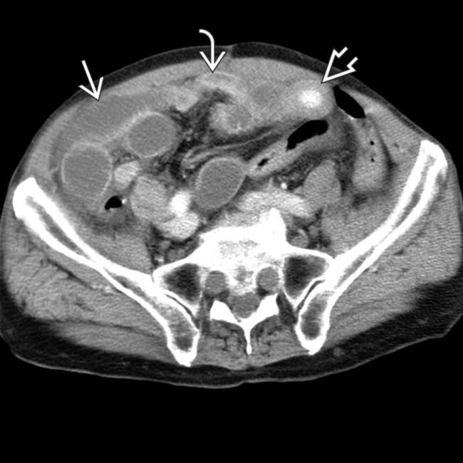
 and decompressed, nondilated distal small bowel
  . The high-density obstructing gallstone
  is seen, as a laminated calcification, at the site of transition from the dilated to the decompressed bowel.
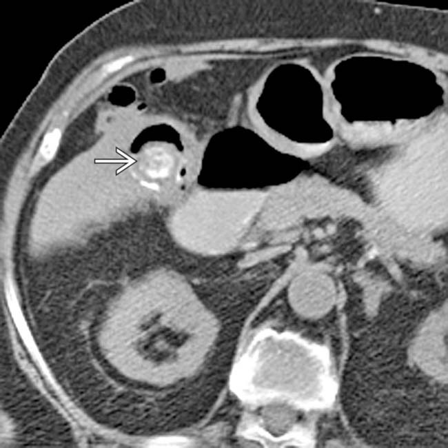
 .
.
 obstructing the distal small bowel.
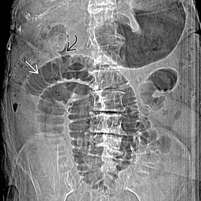
 confirming obstruction, as well as gas in the gallbladder
  .
.
 and laminated filling defect
  at the transition point, which proved to represent an obstructing gallstone.
 and the close proximity of the GB to the duodenum
  .
.
 .
.
 within the dilated small bowel. Note that the bowel distal to the gallstone is collapsed.
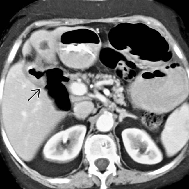
 to the duodenum.
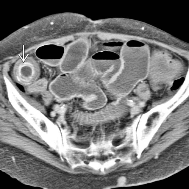
 “stuck” near the ileocecal valve.

