Ultrasound-guided vascular access: Trends and perspectives
Many randomized controlled trials and meta-analyses have associated UGVA with a considerable reduction in complications and increased first-attempt success when compared with standard landmark techniques.1–3 As 1 of their 11 practices to improve patient care, the Agency for Healthcare Research and Quality (AHRQ) recommends the use of ultrasound for placement of a central venous catheter (CVC).4 The National Institute for Clinical Excellence (NICE), as part of their NICE technology guidelines, advocated the routine use of UGVA.5 Most recently, an international, evidence-based consensus statement was developed to assist clinicians in performing UGVA and as a reference for future clinical research. The group advised that ultrasound guidance can be used not only for central venous cannulation but also for peripheral and arterial cannulation. In addition to guidance, the group recommended the use of ultrasound to check for immediate and life-threatening complications, as well as catheter positioning.6
Patient and technical considerations
The implementation of a simple three-step UGVA technique (pre-procedural scanning, real-time ultrasound-guided cannulation, post-procedural scanning) maximizes success rates and decreases complications in the intensive care unit (ICU).6 A pre-procedural scan is recommended to guide the selection of an optimal target vessel.4–6 Usual criteria for the selection of an optimal vein are:
Normal venous patency (venous collapse during breathing or compression without signs of thrombosis)
Easy accessibility (relatively short distance from the skin surface to the vessel wall)
Adequate size (vessel size less than three times the caliber of catheter may carry a greater risk for thrombosis)7
Absence of anatomical variation
Intended purpose of cannulation (i.e., neck surgery often requires infraclavicular vascular access)
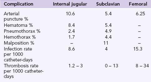
Complication rates after the implementation of landmark techniques differ between sites (Chapter 10). The internal jugular vein carries the highest risk of accidental arterial puncture and hematoma, the subclavian carries the highest risk for pneumothorax, hemothorax, and catheter malposition, while the femoral vein carries the highest risk for thrombosis and infection (Table 11-1). The subclavian vein is considered advantageous in the ICU (compared to the internal jugular or the femoral vein) as it carries the lowest infection risk. However, recent studies show that Chlorhexidine gluconate -impregnated sponge dressing when used with the standard care decreases the incidence of major catheter-related infections from 1.4 to 0.6 per 1000 catheter-days.8 The American Society of Anesthesiologists Task Force on Central Venous Access recommends the use of a central line insertion work and safety checklist and bundling of required equipment to minimize errors, risk of infection, and complications.9 These measures have shown reduction (up to 66%) in central line-associated bloodstream infections.10
Preprocedural tips
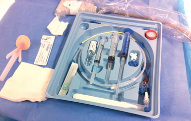
• The implementation of a strict sterilization process including use of a sterile probe cover and gel (Figure 11-1)
• Avoid applying extreme probe pressure on the vessel as normal veins are collapsible vessels
• Optimize the two-dimensional image: center the image on the screen and adjust depth, gain and focus, while obtaining the proper orientation of anatomy with standardization of the dot on the left (Chapter 1).
• When a clear two-dimensional image of the vein is obtained check its patency by applying probe pressure to exclude thrombosis (Video 11-1).
Notably, a thorough preprocedural scan at a prospective region of interest (ROI) should be performed because of the frequent finding of venous and arterial asymmetry between symmetric sites. In that sense, anatomic diversities may exist as well in occasional patients (e.g., duplicated femoral vein). Venous compressibility and patency should be examined because exclusion of thrombosis is mandatory (Chapter 9). The latter is more commonly observed at the common femoral vein site than at other cannulation sites, and ultrasound monitoring of a central line may reveal cases of catheter-related thrombosis (Figure 11-2). Occasionally, trauma to the venous wall, trapped air, hematomas, arteriovenous fistulas, or injuries to nonvascular adjacent structures may be identified after a “clumsy” blind attempt or multiple blind penetrations, which could produce local trauma (Figure 11-3). Estimating vessel size is equally important, as previously mentioned. The size of the internal jugular vein can be evaluated before and after a Valsalva maneuver, which may make a relatively small-caliber vessel that is seemingly difficult to catheterize become robust and easy to puncture.11 Moreover, the diameter of the internal jugular vein is usually found to be larger when the vessel is depicted in the lower neck area (scanning caudally toward the clavicle) than in the upper neck area (scanning cranially toward the mandible). When planning on catheterizing the internal jugular or subclavian vein, the pleura should be assessed for a sliding lung while identifying the vasculature.
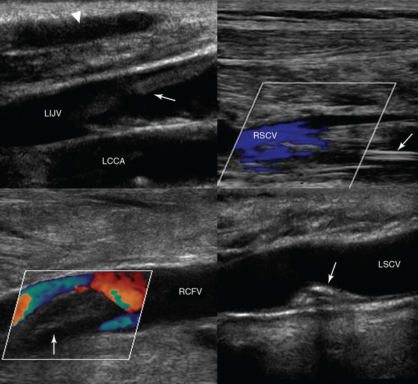
Figure 11-2 (Top) Depiction of a superficial hematoma (arrowhead) and a fresh floating thrombus in the left internal jugular vein (LIJV), which is overlying the left common carotid artery (LCCA), on a longitudinal plane (left). A longitudinal view of the right subclavian vein (RSCV) shows partial flow from central line–associated thrombosis (arrow). (Bottom) Fresh thrombus (arrow) obstructing flow in the right common femoral vein (RCFV) on a longitudinal plane (left). A longitudinal view of the left subclavian vein (LSCV) shows a calcified remnant of a thrombus (following treatment with an anticoagulant) attached to its posterior wall that is not obstructing the vessel’s lumen (right).
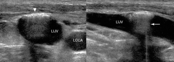
Figure 11-3 Sequelae of a blind attempt at cannulation of the left internal jugular vein (LIJV). LCCA, left common carotid artery. Transverse (left) and longitudinal (right)
Related posts:
 General chest ultrasound in neurocritical care
 Integrating ultrasound into critical care teaching rounds
 Training and competence in ultrasound-guided vascular access
 Ultrasound-guided placement of peripherally inserted central venous catheters
 Lung ultrasound: Protocols in acute dyspnea
 Evaluation of fluid responsiveness by ultrasound
