Transport of Gametes and Fertilization

Published on 13/06/2015 by admin

Filed under Basic Science

Last modified 13/06/2015

Print this page

rate 1 star rate 2 star rate 3 star rate 4 star rate 5 star
Your rating: none, Average: 5 (1 votes)

This article have been viewed 7025 times

Chapter 2

Transport of Gametes and Fertilization

Chapter 1 describes the origins and maturation of male and female gametes and the hormonal conditions that make such maturation possible. It also describes the cyclic, hormonally controlled changes in the female reproductive tract that ready it for fertilization and the support of embryonic development. This chapter first explains the way the egg and sperm cells come together in the female reproductive tract so that fertilization can occur. It then outlines the complex set of interactions involved in fertilization of the egg by a sperm.

Ovulation and Egg and Sperm Transport

Ovulation

Toward the midpoint of the menstrual cycle, the mature graafian follicle, containing the egg that has been arrested in prophase of the first meiotic division, has moved to the surface of the ovary. Under the influence of follicle-stimulating hormone (FSH) and luteinizing hormone (LH), the follicle expands dramatically. The first meiotic division is completed, and the second meiotic division proceeds until the metaphase stage, at which the second meiotic arrest occurs. After the first meiotic division, the first polar body is expelled. By this point, the follicle bulges from the surface of the ovary. The apex of the protrusion is the stigma.

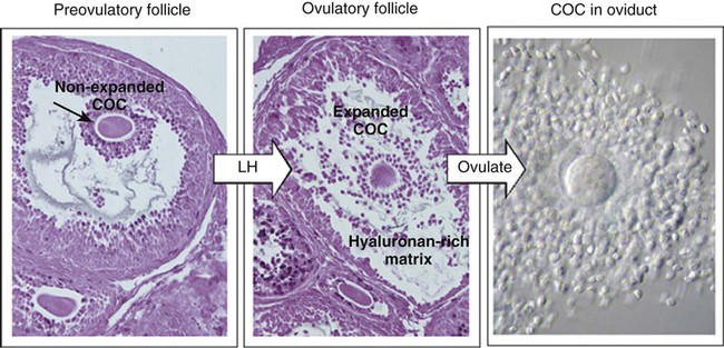
The stimulus for ovulation is the surge of LH secreted by the anterior pituitary at the midpoint of the menstrual cycle (see Fig. 1.16). Within hours of exposure to the LH surge, the follicle reorganizes its program of gene expression from one directed toward development of the follicle to one producing molecules that set into gear the processes of follicular rupture and ovulation. Shortly after the LH peak, local blood flow increases in the outer layers of the follicular wall. Along with the increased blood flow, plasma proteins leak into the tissues through the postcapillary venules, with resulting local edema. The edema and the release of certain pharmacologically active compounds, such as prostaglandins, histamine, vasopressin, and plasminogen activator, provide the starting point for a series of reactions that result in the local production of matrix metalloproteinases—a family of lytic enzymes that degrade components of the extracellular matrix. At the same time, the secretion of hyaluronic acid by cells of the cumulus results in a loosening of the cells surrounding the egg. The lytic action of the matrix metalloproteinases produces an inflammatorylike reaction that ultimately results in rupture of the outer follicular wall about 28 to 36 hours after the LH surge (Fig. 2.1). Within minutes after rupture of the follicular wall, the cumulus oophorus detaches from the granulosa, and the egg is released from the ovary.

Ovulation results in the expulsion of both antral fluid and the ovum from the ovary into the peritoneal cavity. The ovum is not ovulated as a single naked cell, but as a complex consisting of (1) the ovum, (2) the zona pellucida, (3) the two- to three-cell-thick corona radiata, and (4) a sticky matrix containing surrounding cells of the cumulus oophorus. By convention, the adhering cumulus cells are designated the corona radiata after ovulation has occurred. Normally, one egg is released at ovulation. The release and fertilization of two eggs can result in fraternal twinning.

Some women experience mild to pronounced pain at the time of ovulation. Often called mittelschmerz (German for “middle pain”), this pain may accompany slight bleeding from the ruptured follicle.

Egg Transport

The first step in egg transport is capture of the ovulated egg by the uterine tube. Shortly before ovulation, the epithelial cells of the uterine tube become more highly ciliated, and smooth muscle activity in the tube and its suspensory ligament increases as the result of hormonal influences. By ovulation, the fimbriae of the uterine tube move closer to the ovary and seem to sweep rhythmically over its surface. This action, in addition to the currents set up by the cilia, efficiently captures the ovulated egg complex. Experimental studies on rabbits have shown that the bulk provided by the cellular coverings of the ovulated egg is important in facilitating the egg’s capture and transport by the uterine tube. Denuded ova or inert objects of that size are not so readily transported. Capture of the egg by the uterine tube also involves an adhesive interaction between the egg complex and the ciliary surface of the tube.

Even without these types of natural adaptations, the ability of the uterine tubes to capture eggs is remarkable. If the fimbriated end of the tube has been removed, egg capture occurs remarkably often, and pregnancies have even occurred in women who have had one ovary and the contralateral uterine tube removed. In such cases, the ovulated egg would have to travel free in the pelvic cavity for a considerable distance before entering the ostium of the uterine tube on the other side.

When inside the uterine tube, the egg is transported toward the uterus, mainly as the result of contractions of the smooth musculature of the tubal wall. Although the cilia lining the tubal mucosa may also play a role in egg transport, their action is not obligatory because women with immotile cilia syndrome are often fertile.

While in the uterine tube, the egg is bathed in tubal fluid, which is a combination of secretion by the tubal epithelial cells and transudate from capillaries just below the epithelium. In some mammals, exposure to oviductal secretions is important to the survival of the ovum and for modifying the composition of the zona pellucida, but the role of tubal fluid in humans is less clear.

Tubal transport of the egg usually takes 3 to 4 days, whether or not fertilization occurs (Fig. 2.2). Egg transport typically occurs in two phases: slow transport in the ampulla (approximately 72 hours) and a more rapid phase (8 hours) during which the egg or embryo passes through the isthmus and into the uterus (see p. 51). By a poorly understood mechanism, possibly local edema or reduced muscular activity, the egg is temporarily prevented from entering the isthmic portion of the tube, but under the influence of progesterone, the uterotubal junction relaxes and permits entry of the ovum.

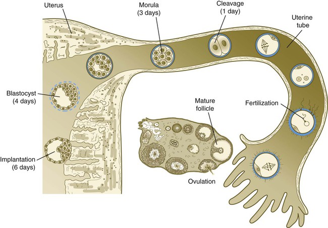
By roughly 80 hours after ovulation, the ovulated egg or embryo has passed from the uterine tube into the uterus. If fertilization has not occurred, the egg degenerates and is phagocytized. (Implantation of the embryo is discussed in Chapter 3.)

Sperm Transport

Sperm transport occurs in both the male reproductive tract and the female reproductive tract. In the male reproductive tract, transport of spermatozoa is closely connected with their structural and functional maturation, whereas in the female reproductive tract, it is important for spermatozoa to pass to the upper uterine tube, where they can meet the ovulated egg.

After spermiogenesis in the seminiferous tubules, the spermatozoa are morphologically mature but are nonmotile and incapable of fertilizing an egg (Fig. 2.3). Spermatozoa are passively transported via testicular fluid from the seminiferous tubules to the caput (head) of the epididymis through the rete testis and the efferent ductules. They are propelled by fluid pressure generated in the seminiferous tubules and are assisted by smooth muscle contractions and ciliary currents in the efferent ductules. Spermatozoa spend about 12 days in the highly convoluted duct of the epididymis, which measures 6 m in the human, during which time they undergo biochemical maturation. This period of maturation is associated with changes in the glycoproteins in the plasma membrane of the sperm head. By the time the spermatozoa have reached the cauda (tail) of the epididymis, they are capable of fertilizing an egg.

On ejaculation, the spermatozoa rapidly pass through the ductus deferens and become mixed with fluid secretions from the seminal vesicles and prostate gland. Prostatic fluid is rich in citric acid, acid phosphatase, zinc, and magnesium ions, whereas fluid of the seminal vesicle is rich in fructose (the principal energy source of spermatozoa) and prostaglandins. The 2 to 6 mL of ejaculate (semen, or seminal fluid) typically consists of 40 to 250 million spermatozoa mixed with alkaline fluid from the seminal vesicles (60% of the total) and acid secretion (pH 6.5) from the prostate (30% of the total). The pH of normal semen ranges from 7.2 to 7.8. Despite the numerous spermatozoa (>100 million) normally present in an ejaculate, a number as small as 25 million spermatozoa per ejaculate may be compatible with fertility.

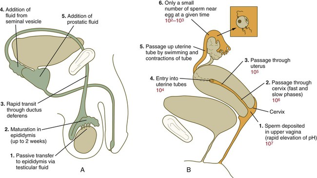
In the female reproductive tract, sperm transport begins in the upper vagina and ends in the ampulla of the uterine tube, where the spermatozoa make contact with the ovulated egg. During copulation, the seminal fluid is normally deposited in the upper vagina (see Fig. 2.3), where its composition and buffering capacity immediately protect the spermatozoa from the acid fluid found in the upper vaginal area. The acidic vaginal fluid normally serves a bactericidal function in protecting the cervical canal from pathogenic organisms. Within about 10 seconds, the pH of the upper vagina is increased from 4.3 to as much as 7.2. The buffering effect lasts only a few minutes in humans, but it provides enough time for the spermatozoa to approach the cervix in an environment (pH 6.0 to 6.5) optimal for sperm motility.

The next barriers that the sperm cells must overcome are the cervical canal and the cervical mucus that blocks it. Changes in intravaginal pressure may suck spermatozoa into the cervical os, but swimming movements also seem to be important for most spermatozoa in penetrating the cervical mucus.

The composition and viscosity of cervical mucus vary considerably throughout the menstrual cycle. Composed of cervical mucin (a glycoprotein with a high carbohydrate composition) and soluble components, cervical mucus is not readily penetrable. Between days 9 and 16 of the cycle, however, its water content increases, and this change facilitates the passage of sperm through the cervix around the time of ovulation; such mucus is sometimes called E mucus. After ovulation, under the influence of progesterone, the production of watery cervical mucus ceases, and a new type of sticky mucus, which has a much decreased water content, is produced. This progestational mucus, sometimes called G mucus, is almost completely resistant to sperm penetration. A highly effective method of natural family planning makes use of the properties of cervical mucus.

There are two main modes of sperm transport through the cervix. One is a phase of initial rapid transport, by which some spermatozoa can reach the uterine tubes within 5 to 20 minutes of ejaculation. Such rapid transport relies more on muscular movements of the female reproductive tract than on the motility of the spermatozoa themselves. These early-arriving sperm, however, appear not to be as capable of fertilizing an egg as do those that have spent more time in the female reproductive tract. The second, slow phase of sperm transport involves the swimming of spermatozoa through the cervical mucus (traveling at a rate of 2 to 3 mm/hour), their storage in cervical crypts, and their final passage through the cervical canal as much as 2 to 4 days later.

Relatively little is known about the passage of spermatozoa through the uterine cavity, but the contraction of uterine smooth muscle, rather than sperm motility, seems to be the main intrauterine transport mechanism. At this point, the spermatozoa enter one of the uterine tubes. According to some more recent estimates, only several hundred spermatozoa enter the uterine tubes, and most enter the tube containing the ovulated egg.

Once inside the uterine tube, the spermatozoa collect in the isthmus and bind to the epithelium for about 24 hours. During this time, they are influenced by secretions of the tube to undergo the capacitation reaction. One phase of capacitation is removal of cholesterol from the surface of the sperm. Cholesterol is a component of semen and acts to inhibit premature capacitation. The next phase of capacitation consists of removal of many of the glycoproteins that were deposited on the surface of the spermatozoa during their tenure in the epididymis. Capacitation is required for spermatozoa to be able to fertilize an egg (specifically, to undergo the acrosome reaction; see p. 29). After the capacitation reaction, the spermatozoa undergo a period of hyperactivity and detach from the tubal epithelium. Hyperactivation helps the spermatozoa to break free of the bonds that held them to the tubal epithelium. It also assists the sperm in penetrating isthmic mucus, as well as the corona radiata and the zona pellucida, which surround the ovum. Only small numbers of sperm are released at a given time. This may reduce the chances of polyspermy (see p. 31).

On their release from the isthmus, the spermatozoa make their way up the tube through a combination of muscular movements of the tube and some swimming movements. The simultaneous transport of an egg down and spermatozoa up the tube is currently explained on the basis of peristaltic contractions of the uterine tube muscles. These contractions subdivide the tube into compartments. Within a given compartment, the gametes are caught up in churning movements that over 1 or 2 days bring the egg and spermatozoa together. Fertilization of the egg normally occurs in the ampullary portion (upper third) of the uterine tube. Estimates suggest that spermatozoa retain their function in the female reproductive tract for about 80 hours.

After years of debate concerning the possibility that mammalian spermatozoa may be guided to the egg through attractants, more recent research suggests that this could be the case. Mammalian spermatozoa have been found to possess odorant receptors of the same family as olfactory receptors in the nose, and they can respond behaviorally to chemically defined odorants. Human spermatozoa also respond to cumulus-derived progesterone and to yet undefined chemoattractants emanating from follicular fluid and cumulus cells. Human spermatozoa are also known to respond to a temperature gradient, and studies on rabbits have shown that the site of sperm storage in the oviduct is cooler than that farther up the tube where fertilization occurs. It seems that only capacitated spermatozoa have the capability of responding to chemical or thermal stimuli. Because many of the sperm cells that enter the uterine tube fail to become capacitated, these spermatozoa are less likely to find their way to the egg.

Formation and Function of the Corpus Luteum of Ovulation and Pregnancy

While the ovulated egg is passing through the uterine tubes, the ruptured follicle from which it arose undergoes a series of striking changes that are essential for the progression of events leading to and supporting pregnancy (see Fig. 1.8). Soon after ovulation, the basement membrane that separates the granulosa cells from the theca interna breaks down, thus allowing thecal blood vessels to grow into the cavity of the ruptured follicle. The granulosa cells simultaneously undergo a series of major changes in form and function (luteinization). Within 30 to 40 hours of the LH surge, these cells, now called granulosa lutein cells, begin secreting increasing amounts of progesterone along with some estrogen. This pattern of secretion provides the hormonal basis for the changes in the female reproductive tissues during the last half of the menstrual cycle. During this period, the follicle continues to enlarge. Because of its yellow color, it is known as the corpus luteum. The granulosa lutein cells are terminally differentiated. They have stopped dividing, but they continue to secrete progesterone for 10 days.

In the absence of fertilization and a hormonal stimulus provided by the early embryo, the corpus luteum begins to deteriorate (luteolysis) late in the menstrual cycle. Luteolysis seems to involve both the preprogramming of the luteal cells to apoptosis (cell death) and uterine luteolytic factors, such as prostaglandin F2. Regression of the corpus luteum and the accompanying reduction in progesterone production cause the hormonal withdrawal that results in the degenerative changes of the endometrial tissue during the last days of the menstrual cycle.

During the regression of the corpus luteum, the granulosa lutein cells degenerate and are replaced with collagenous scar tissue. Because of its white color, the former corpus luteum now becomes known as the corpus albicans (“white body”).

If fertilization occurs, the production of the protein hormone chorionic gonadotropin by the future placental tissues maintains the corpus luteum in a functional condition and causes an increase in its size and hormone production. Because the granulosa lutein cells are unable to divide and cease producing progesterone after 10 days, the large corpus luteum of pregnancy is composed principally of theca lutein cells. The corpus luteum of pregnancy remains functional for the first few months of pregnancy. After the second month, the placenta produces enough estrogens and progesterone to maintain pregnancy on its own. At this point, the ovaries can be removed, and pregnancy would continue.

Fertilization

Fertilization is a series of processes rather than a single event. Viewed in the broadest sense, these processes begin when spermatozoa start to penetrate the corona radiata that surrounds the egg and end with the intermingling of the maternal and paternal chromosomes after the spermatozoon has entered the egg.

Penetration of the Corona Radiata

When the spermatozoa first encounter the ovulated egg in the ampullary part of the uterine tube, they are confronted by the corona radiata and some remnants of the cumulus oophorus, which represents the outer layer of the egg complex (Fig. 2.4). The corona radiata is a highly cellular layer with an intercellular matrix consisting of proteins and a high concentration of carbohydrates, especially hyaluronic acid. It is widely believed that hyaluronidase emanating from the sperm head plays a major role in penetration of the corona radiata, but the active swimming movements of the spermatozoa are also important.
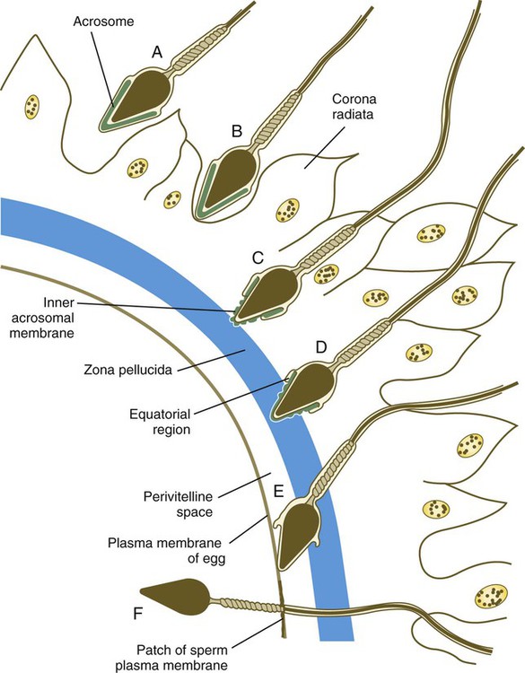
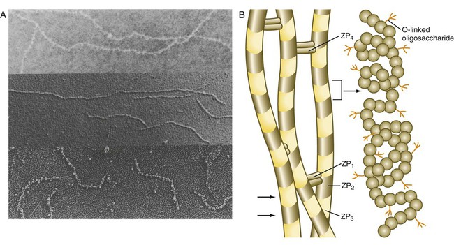