The maxillary antra
Introduction
The maxillary antra, because of their close proximity to the upper teeth, are the most important of the paranasal sinuses in dentistry. Nowadays the imaging modalities of choice to investigate possible disease within the antra are computed tomography (CT), cone beam CT (CBCT) and magnetic resonance (MR) (see Chapters 16 and 18). However, as the antra are often imaged on conventional dental radiographs, as well as on certain skull views, clinicians still need to know:
• The anatomy of the antra, including their shape, size, normal variations and related structures.
• How the antra are represented normally on conventional dental and skull radiographs and how these radiographs are assessed.
Similar radiographic changes can be seen in the other paranasal sinuses – frontal, ethmoidal and sphenoidal. They are of less clinical relevance in dentistry and are discussed only briefly.
Normal anatomy
The main anatomical parts of the antra (see Fig. 31.1) can be divided into:
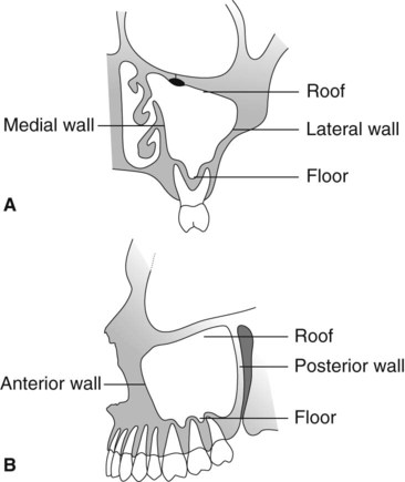
• A roof or upper border, bounded by the orbit
• A medial wall, bounded by the nasal cavity
• A posterior wall, related to the pterygopalatine fossa
• A lateral wall, related to the zygoma and cheek
• An anterior wall, related to the cheek
• A floor, related to the apices of the upper posterior teeth.
Normal appearance of the antra on conventional radiographs
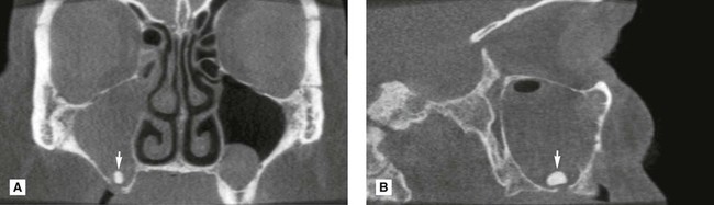
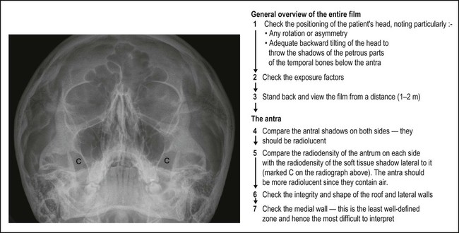
An antrum appears radiographically as a radiolucent cavity in the maxilla, with well-defined, dense, corticated radiopaque margins or walls. In general, the larger the cavity the more radiolucent it will appear. The internal bony septa and blood vessel canals in the walls all produce their own shadows. The thin epithelial lining is not normally seen. The different parts of the antra shown on conventional dental and skull radiographs are summarized in Table 31.1. Typical normal radiographic appearances are shown in Figs 31.2–31.4. In addition, a suggested systematic approach to viewing the antra on the 0° OM is shown in Fig. 31.4.
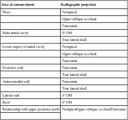
Table 31.1
Summary of the different parts of the antra shown on conventional dental and skull radiographs
| Area of antrum shown | Radiographic projection |
| Floor | Periapical |
| Upper oblique occlusal | |
| Panoramic | |
| Main antral cavity | 0° OM |
| True lateral skull | |
| Lower aspect of antral cavity | Periapical |
| Upper oblique occlusal | |
| Panoramic | |
| Posterior wall | Panoramic |
| True lateral skull | |
| Anteromedial wall | Panoramic |
| True lateral skull | |
| Lateral wall | 0° OM |
| Roof | 0° OM |
| Relationship with upper posterior teeth | PeriapicalUpper oblique occlusalPanoramic |
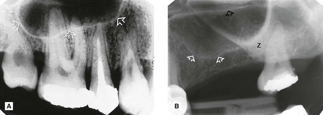
 showing the usual appearance of the floor (arrowed) and base of the antral cavity in relation to the upper posterior teeth in a dentate adult. B Periapical of
  of a partially dentate adult showing various normal anatomical structures. These include the floor of the antrum (open white arrows), the hard palate/floor of the nasal cavity (open black arrows) and the zygomatic buttress (Z).
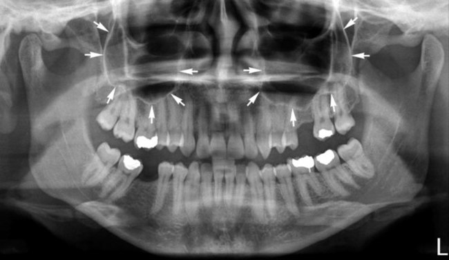
Antral disease
The major pathological conditions that can affect the antra, directly or indirectly, include:
These various disease entities can result in the following radiological changes:
• Total or partial opacity/obliteration of the main antral cavity
• Alteration in the integrity of the antral walls, including discontinuity as a result of a fracture or destruction by an intrinsic or extrinsic tumour
• Alteration in the antral outline, including expansion or compression caused by an intrinsic or extrinsic lesion or disease process
Investigation and appearance of disease within the antra
• Computed tomography (CT) – currently recommended by the Royal College of Radiologists in the UK
• Multidirectional (spiral) tomography
Appearances of antral diseases using various imaging modalities are shown in Figs 31.5–31.23.