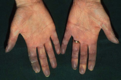
Raynaud disease and phenomenon
Sameh S. Zaghloul, Najat A.Y. Marraiki and Mark J.D. Goodfield
Specific investigations


First-line therapies
 Calcium channel blockers
 Glyceryl trinitrate
 Prostacyclin analogs
Buy Membership for Dermatology Category to continue reading. Learn more here
Sameh S. Zaghloul, Najat A.Y. Marraiki and Mark J.D. Goodfield
