Radiotherapy for Head and Neck Cancer: Radiation Physics, Radiobiology, and Clinical Principles

Published on 03/05/2015 by admin

Filed under Otolaryngology

Last modified 03/05/2015

Print this page

rate 1 star rate 2 star rate 3 star rate 4 star rate 5 star
Your rating: none, Average: 4.5 (2 votes)

This article have been viewed 3503 times

CHAPTER 77 Radiotherapy for Head and Neck Cancer

Radiation Physics, Radiobiology, and Clinical Principles

Key Points

Basic Radiation Physics

Radiation Biology

Clinical Principles of Radiotherapy

The aim of this chapter is to provide an outline of the basic principles of physics and biophysiology that lie behind modern radiotherapy. This overview is intended to enable the clinician to understand the physics background of the various radiation treatments, with a special focus on head and neck treatment technologies.

Toward the closing years of the nineteenth century, many investigations into electricity characteristics were conducted, one of them demonstrating that electric potential placed across two separated platinum electrodes produces a spark.1 British physicist William Crookes demonstrated that if the two electrodes were placed within an evacuated glass tube, the vacuum would eventually cause the walls of the vessel to fluoresce.2 On November 8, 1895, Wilhelm Roentgen, while performing an experiment with the Crookes tube, accidentally left a piece of paper painted with barium platinocyanide nearby; he noted that the paper fluoresced and realized that this fluorescence of the paper could have been caused by a new, invisible type of ray that the tube was now emitting. Thus, the x-ray was discovered.

Radioactivity was discovered shortly after by Henri Becquerel, who investigated the capability of different substances to produce x-rays. He observed the darkening of photographic plates by uranium salts and concluded that the same x-rays were emitted spontaneously and continuously from the uranium.3 Pierre and Marie Curie, who read his results, coined the term radioactivity to describe this phenomenon. In 1898 they isolated a material with radioactivity 60 times higher than uranium and called it radium.4

These discoveries led to radiation biology experiments. The first documented experiment was performed unintentionally at about the same time, when Antoine Becquerel developed a “burn” on his chest from carrying a vial of radium salt in his vest pocket. It soon became apparent that radiation had the ability to produce profound biologic changes. In the beginning it was believed to be a magical cure for almost every known illness. The first documented success was reported in 1899 in Stockholm5 by Thor Stenbeck, who treated a 49-year-old woman’s nasal basal-cell carcinoma. He delivered 100 treatments in the course of 9 months, and the patient was alive and well 30 years after the treatment. In 1901, Dr. Frand Williams in Boston reported on the successful treatment of a lip cancer.5 The early treatments often involved very large single exposures that resulted in extensive skin toxicities and other complications. Therefore, only superficial sites were originally treated by the direct application of radium.6 Eventually, physicians started to insert radium directly into deep-seated tumors, effectively beginning the field of brachytherapy.7

The use of external beam treatments made its leap in 1922 when Coutard and Hautant reported a new concept of fractionated treatments: advanced laryngeal cancer could be cured without severe toxicities using fractionated treatments.8 Advances in measurements were also achieved when the skin erythema dose (the dose of x-rays required to give a light skin reaction) was replaced by the roentgen in 1928,9 which was later replaced by the rad. The rad is the unit of absorbed dose and is a measure of the energy deposition per unit mass by all types of ionizing radiation. The next step was achieved with the development of higher-energy machines capable of depositing dose at depth.

As technology has progressed, radiation therapy (RT) has become increasingly sophisticated, with computer controls to deliver exact and modulated doses to depths and specific areas within the treatment field. The advances in energy technologies led to the routine use of high-energy and accurate deep-penetrating radiation produced by linear accelerators. Nuclear physics innovations produced many artificial radioisotopes, enabling the use of high-dose brachytherapy, which shortens treatment time and simplifies the radioprotection procedures.

Aside from the technology advances, radiation treatment has a special role in head and neck cancer (HNC), in which it has in many cases a major advantage over traditional surgery in its ability to preserve organs, improving quality of life without compromising the survival rate.

Basic Physics

Characteristics of Radiation

Radiation refers to the propagation of energy through space or a medium (Fig. 77-1). Radiation can be broadly classified as either particulate or electromagnetic. If the radiant energy is carried off by a particle that has rest mass, the radiation is called “corpuscular” or “particulate” radiation. Examples of particulate radiations are electrons, beta particles, protons, neutrons, and heavy charged particles.

image

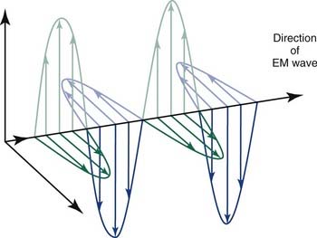
Figure 77-1. Propagation of electromagnetic (EM) radiation with oscillating electric and magnetic fields.

(From Saw CB. Foundation of Radiological Physics. Omaha, NE: C. B. Saw Publishing; 2004:10, with permission.)

Electron beams, which are produced in a linear accelerator, are widely available for the treatment of superficial lesions in clinical radiation oncology practice. In addition, mixed beams consisting of electron beams and photon beams are used to treat lesions that require higher surface doses. Negatrons from strontium Sr-90 are used in the treatment of restenosis in intravascular coronary brachytherapy. Although other forms of particulate radiation have been used experimentally in the past, their use has been limited to a few centers. The use of protons is gaining popularity in many centers specifically for pediatric tumors.

Electromagnetic radiation is a packet of energy (a photon) that propagates through space. It has no rest mass and propagates at the speed of light. This discrete energy (E) is related to its associated frequency (ν) as follows:

(Eq. 77-1) image

where h is Planck’s constant, having a value of 6.626 × 10⁻³⁴ joule-second (J.s). The energy of a photon is often expressed in electron volts (eV). One eV represents the amount of energy required to accelerate an electron through a potential of one volt. The frequency (ν) of a photon is related to its wavelength (λ) as follows:

(Eq. 77-2) image

where c = 3.0 × 10⁸ m/sec, the speed of light in a vacuum. The waves of electromagnetic radiation are composed of oscillating electric and magnetic fields that are orthogonal to each other and to the direction of propagation, as shown in Figure 77-1. The electromagnetic spectrum spans a broad and continuous range from radiowaves to x-rays with wavelengths from 106 to 10−13 m. Radiation with wavelengths shorter than visible light is classified as ultraviolet rays, x-rays, and gamma rays. The boundaries between these regions are not sharply defined. For example, x-rays and gamma rays are indistinguishable except for their origins, one from the orbital electrons and the other from the nucleus, respectively. Different types of electromagnetic radiation interact differently with the same material (see Fig. 77-1).

Radiation Production from Linear Accelerators

Another type of radiation is termed bremsstrahlung11 (German for “braking radiation”), electromagnetic radiation produced by a sudden slowing down or deflection of charged particles (especially electrons) passing through matter near the strong electric fields of atomic nuclei.

Internal bremsstrahlung arises in the radioactive process of beta decay, which consists of the production and emission of electrons by unstable atomic nuclei or the capture by nuclei of one of their own orbiting electrons. These electrons, deflected from their own associated nuclei, emit internal bremsstrahlung. During the deceleration process, the electrons interact with the atomic nuclei, resulting in the emission of bremsstrahlung radiation. Before 1950, external beam RT was accomplished in this way, with a maximum energy of about 300 keV. As just stated, these x-rays are low in energy compared to what is used today, with the disadvantages of poor penetration and maximal deposition of dose at the skin.

Today, other types of radiation are produced in nuclear reactors, cyclotrons, and linear accelerators.

Interaction of X-Rays with Matter

After these high-energy x-rays are produced successfully, they can interact with matter via several different processes. Each interaction type has a probability, based on the composition of the matter and the energy of the x-rays. These interactions cause some photons (x-rays) to be removed from the forward-moving x-ray beam, an effect called attenuation, which is basically the loss of intensity and subsequent decrease in the deposition of dose as the beam reaches greater depths. The five possible interactions of x-rays with matter are as follows:

The most important of these interactions in RT are represented in Figure 77-2.

image

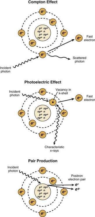
Figure 77-2. The major radiation interactions. e, electron; n, neutron; p, proton.

(Redrawn from Cox JD, Ang KK, eds. Radiation Oncology: Rationale, Technique, Results. 8th ed. St. Louis: Mosby; 2003:5.)

Deposition of Dose

The absorbed dose from an x-ray beam is the measure of the energy deposited by the beam and absorbed by the target. Radiation doses were historically measured by roentgen, which was a unit of exposure but did not quantify the dose the patient absorbs. In the late 1950s the rad, an abbreviation for “radiation-absorbed dose,” was introduced. The rad is equivalent to the deposition of 100 ergs (10−7 joules) per gram of material.

More recently, an international commission has agreed that radiation doses should be specified in terms of gray (Gy), which corresponds to the deposition of 1 joule per kilogram of material. Numerically, doses in rad can be converted to equivalent doses in Gy by dividing by 100 (i.e., 100 rad = 1 Gy). This is closely related to the observed biologic effects, how and where the dose is deposited is obviously very important. Because the amount of radiation absorbed by the target is assumed to be as stated previously, x-rays in the megavoltage energy range, such as those used in RT, exhibit the phenomenon of skin sparing, whereby the dose deposited in tissue is relatively low at the surface but increases rapidly over the first few millimeters. The region of rapidly increasing dose is known as the buildup region. This rapid increase occurs because of the forward-moving photons interacting with electrons of the target tissue via the interactions described previously. Because these electrons are also propelled forward but have a shorter course than the photons, there becomes an area at depth at which the number of electrons entering the plane of interaction from superficial interactions is exactly equal to the amount leaving the plane from interactions in that plane. This plane is termed the Dmax, as it represents the maximum number of ionization events past this point; as more interactions between the photon beam and tissue occur, fewer photons are available to travel forward and deposit dose at greater depths. This process, as previously described, is called attenuation. How quickly a photon beam is attenuated (i.e., how much dose it deposits at depth) depends on qualities both within the beam and within the target tissue. The most substantial effect on depth dose from the photon beam itself is the beam’s energy.

Deposition of Energy

As a high-energy photon beam traverses through a medium, the atoms are excited and ionized. Of the two interacting processes, ionization is disruptive to atoms, changing the molecular integrity and leading to cell death in tissue. The ejected electrons undergo further interactions and deposit their energies to the medium through excitation and ionization. The amount of energy transferred to the electrons at the initial point of interaction is called kerma. The second stage of the energy-transfer process is called the absorbed dose. Not all the energy transferred to the medium at the initial point of interaction is absorbed. Some of the energy is radiated away as bremsstrahlung, and the remaining is called collision kerma. The absorbed dose is the energy retained in the medium along the path of the electrons. If the electron track has an appreciable length, the transfer of energy (kerma) and the absorption of energy (absorbed dose) take place at separate locations.

When a photon beam enters a medium, the initial ionization of atoms takes place at the surface. The energy is directly transferred to the ejected electrons, and therefore kerma has a maximum value at the surface. As the beam proceeds farther, the photon intensity decreases as a result of absorption and scatter. Hence kerma decreases as a function of depth into the medium. On the other hand, absorbed dose is initially low at the medium surface, increases to a maximum, and thereafter decreases as a function of depth. The region from the surface to depth of maximum dose is called the buildup region. The position of maximum absorbed dose is called the point of equilibrium, and the region beyond the maximum dose is the region of transient electronic equilibrium.

Radiation in Conventional Medicine

External beam radiotherapy is the most frequently used form of radiotherapy. With this technique, the patient lies on a bed and an external source of radiation is pointed at a particular part of the body. Kilovoltage (which may be either superficial or deep) x-rays are used for treating skin cancer and superficial structures. Megavoltage x-rays are used to treat deep-seated tumors (e.g., bladder, bowel, prostate, lung, brain). Megavoltage electrons are used mainly for treating superficial tumors.

The energy of diagnostic and therapeutic gamma rays and x-rays is expressed in kilovolts (kV)or megavolts (MV), whereas the energy of therapeutic electrons is expressed in terms of megaelectron volts (MeV). In diagnostic and therapeutic situations, this voltage is the maximum electric potential used by a linear accelerator to produce the photon beam. The beam is made up of a spectrum of energies: the maximum energy is approximately equal to the beam’s maximum electric potential times the electron charge. Thus, a 1-MV beam will produce photons of no more than about 1 MeV. The mean x-ray energy is only about a third of the maximum energy. Beam quality and hardness may be improved by special filters, which improve the homogeneity of the x-ray spectrum.

In the medical field, useful x-rays are produced when electrons are accelerated to a high energy. Some examples of x-ray energies used in medicine are as follows:

Of these energy ranges, megavoltage x-rays are by far the most common in radiotherapy. Orthovoltage x-rays do have limited applications, and the other energy ranges are not typically used clinically.

Medically useful photon beams can also be derived from a radioactive source such as cobalt Co-60, iridium Ir-192, cesium Cs-137 and radium Ra-226 (which is no longer used clinically). Such photon beams, derived from radioactive decay, are more or less monochromatic and are properly termed gamma rays.

Orthovoltage x-rays are produced by linear accelerators operating at 200 to 500 kV. These are also known as “deep” or “superficial” machines, depending on their energy range. Orthovoltage units have essentially the same design as diagnostic x-ray machines. These machines are generally limited to less than 600 kV.

Linear accelerators (linacs) produce megavoltage x-rays. Commercially available medical linear accelerators produce x-rays and electrons with an energy range from 4 MeV up to around 25 MeV.11 The x-rays themselves are produced by the rapid deceleration of electrons in a target material, typically a tungsten alloy, which produces an x-ray spectrum via bremsstrahlung radiation. The shape and intensity of the beam produced by a linear accelerator may be modified or collimated by a variety of means. Thus, conventional, conformal, intensity-modulated, tomographic, and stereotactic radiotherapy are all produced by specially modified linear accelerators (Fig. 77-3).

image

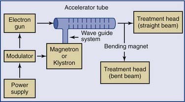
Figure 77-3. Block diagram of a typical medical linear accelerator.

(From Leibel SA, Phillips TL, eds. Textbook of Radiation Oncology. Philadelphia: WB Saunders; 1998:110.)

As the particle bundle passes through the tube it is unaffected, and the frequency of the driving signal and the spacing of the gaps between electrodes is designed so that the maximum voltage differential appears as the particle crosses the gap. This accelerates the particle, imparting energy to it in the form of increased velocity. At speeds near the speed of light the incremental velocity increase will be small, with the energy appearing as an increase in the mass of the particles

Additional magnetic or electrostatic lens elements may be included to ensure that the beam remains in the center of the tube and its electrodes (see Fig. 77-3).

Linear Accelerators

Linear accelerators typically produce beam energies ranging from 6 to 18 MeV, and the dose at depth increases with beam energy. Therefore, an 18-MeV photon beam would deliver more dose to a given depth in a patient than would a 6-MeV photon beam. An 18-MeV beam would also have more skin sparing (i.e., it would have a greater Dmax). Another aspect that affects the depth dose is the size of the field of radiation used to treat the patient. With a larger field size, there is greater scattering of photons within the field during the interactions with electrons. This scatter effect leads to more interactions, which translate into a higher deposition of dose at depth. In other words, the dose at 10-cm depth within a patient from a photon beam that has a field size of 20 cm × 20 cm would be higher than the same photon beam with a field size of 5 cm × 5 cm. Many other factors go into the calculation of dose delivered at varying depths in a patient, including scatter from the collimators in the machine, blocks to shield normal tissue, and wedges and compensators (which are used to shape the photon beam). Another main modifier in the target tissue that affects dose at depth is the density of the tissue being treated. Lung, for example, being less dense than soft tissue, allows more photon transmission. Additionally, the inverse square effect, first noted by Roentgen, must be taken into account. All of these factors must be taken into consideration in the determination of the dose being delivered to structures within the patient. Calculation of the dose given to a tumor or other volumes within a patient is thus complex, requiring much more knowledge than simply how much x-ray dose the machine is putting out.

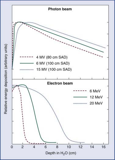
As an energy source, electrons differ from photons in that electrons travel only a certain (short) distance within tissue. They are very light particles compared with the nuclei of the target tissue with which they interact. Hence, the electrons lose a large fraction of their energy in a single process. This leads to much less skin sparing and the deposition of the majority of the dose in superficial tissues. Consequently, however, they are very useful for treatments in which the target of the radiation lies close to the surface of the patient, such as skin tumors (Fig. 77-4).

Particle Beams

High-energy charged particle beams interact with tissues in a unique way. At first, the charged particles lose energy gradually, but there is an intense release of energy at the end of the range. This intense energy deposition is called the Bragg peak. This feature provides a means of delivering the maximum dose to a target at a specified depth. Often in clinical practice, the particle beams are modulated to change the energy, and hence the depth, to widen the dose deposition to cover the target, and this change is in addition to allowing optimized dose delivery.

In the strictest sense, the electron beams used in conventional radiotherapy facilities are a type of particle radiation, but this section is devoted to the heavier charged particles (e.g., protons, α-particles, heavy ions, π-mesons, and fast neutrons) used experimentally at a small number of radiotherapy centers throughout the world. These particles are of special interest because of their different radiobiologic properties or their better depth-dose characteristics, which allow for higher tumor doses without causing a commensurate increase in the dose to the surrounding healthy tissues.

One particle for which there has been a great amount of clinical work is the fast neutron.12,13 Fast neutrons are of clinical interest because of their radiobiologic properties, which occur because of the much greater amount of energy they deposit when they go through tissue. Neutrons are neutral particles and interact with the atomic nuclei, producing “heavy” charged particles such as protons, α-particles, or nuclear fragments that in turn create a dense chain of ionization events as they go through tissue. The distribution of these secondary particles depends on the energy spectrum of the neutron beam, so the biologic properties of the beam depend strongly on its energy spectrum. Neutrons used in therapy generally are produced by accelerating charged particles, such as protons or deuterons, and impacting them on a beryllium target.

There is considerable interest in using charged particle beams directly for therapeutic purposes, which generally requires beams of much higher energy than those used to produce neutrons. The lighter particles, such as protons14 and α-particles, are of interest because of their extremely favorable depth-dose characteristics. The radiobiologic properties of these beams are similar to those of conventional photon or electron beams. Heavy charged particles combine the favorable depth-dose properties of the proton and α-particle beams with the favorable biologic properties of the neutron beams. Energies are on the order of several hundred MeV per nucleon, rather than the few MeV per nucleon for the recoil fragments produced by neutrons. These highly energetic particles do not deposit much energy in tissue until they reach the end of their path, where they are moving slowly. Hence, they do not produce much radiation damage in the intervening tissues. Because of their extremely high cost and general unavailability, however, these beams are not widely in use. Interest in proton beams has grown, and the number of radiotherapy facilities using protons in the United States is expected to rise.

Treatment Aspects of External Beam Radiation

A series of technical tasks must be performed to prepare a patient to undergo external beam RT. The purpose of these tasks—simulation, treatment planning, verification, dose delivery, and quality assurance—is to ensure that high radiation doses are delivered to the patient in an accurate and controlled manner. Usually the processes of simulation, treatment planning, and verification take 1 to 3 days. The setup and treatment planning must be individualized to maximize the dose delivered to the target while minimizing the irradiation of surrounding normal tissue. Any inaccuracy in this process could cause further delay in the initiation of the radiation treatment.

During simulation, the patient setup is assessed for ease of positioning and daily reproducibility. This process involves the use of immobilization devices (anything that can hold a patient in position during treatment) and treatment aids (any kind of support to make patient comfortable). In most HNC cases, a mask that immobilizes the head and neck is used.

After the patient is immobilized, at least two reference points are marked on the patient with x-ray markers to define a reference plane close to the internal tumor target. The marking of the reference points is aided by the use of lasers. Next, the region of interest on the patient is scanned using computed tomography (CT), often using a dedicated CT-simulator scanner to obtain CT images for image-based treatment planning. The images are scanned to a treatment planning station. The radiation oncologist then outlines the tumor target region and critical surrounding normal structures on the axial images on the basis of the CT scan, and a prescribed dose with appropriate normal tissue margin is provided. On the basis of this information, a medical physicist or a medical dosimetrist designs an individualized treatment plan. The challenge in the plan is to deliver the prescribed dose uniformly to the tumor or tumor bed target while maintaining a very low dose to the critical normal structures. Although these goals are achievable, it takes time to arrive at an optimized individualized plan.

Aside from delivering the prescribed dose to the target and minimal dose to the surrounding critical structures, the individualized plan must be evaluated for deliverability—that is, the selected beam orientation must be such that the linear accelerator does not collide with the patient or the table, and the technical parameters of the patient’s plan must comply with the technical requirements of the delivery machine. Next, the individualized machine-delivery parameters are downloaded into a record verification system database. Patient verification is the process of ascertaining that the individualized plan is correct and deliverable. Correct here means that the patient is easily placed into treatment position, relatively comfortable, and immobile, the setup is reproducible, and the target is at the appropriate position relative to the isocenter according to the plan. Finally, there is clearance to avoid patient-equipment collision. During verification, orthogonal radiographs or individualized field radiographs are taken, with films or a portal imager, for assessment and for documentation of the setup. In addition, machine parameters are captured into the database. These images can be used to assist in repositioning the patient as necessary.

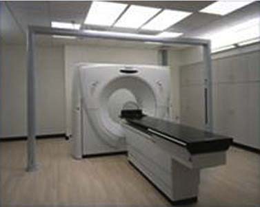
Typically, the radiation dose is delivered to the patient using a linear accelerator (Fig. 77-5). The radiation beam as produced is a forward peak; that is, it has extremely high intensity along the beam axis. It must pass through a conical metallic flattening filter to create a uniform field beam for clinical use. As the clinical beam exits the linear accelerator, it is collimated by a pair of jaws or a multileaf collimation (MLC) system (see Fig. 77-5). The multileaf collimation system is used to shield and protect normal structures (replacing the antiquated and cumbersome lead blocks). In addition, the system is used to modulate beam intensity in intensity-modulated RT. The linear accelerator has a gantry that allows the rotation of the treatment head around the patient. Hence, the radiation beam can be directed at the target from multiple positions to reduce dose to the normal tissues. Because of the high voltage, moving parts, and high dose, the linear accelerator must be properly calibrated and properly maintained for its safe use (see Fig. 77-5).

In addition to external beam therapy, radiation oncology departments offer brachytherapy services. In brachytherapy (brachy is derived from the Greek word for “short”), sealed radioactive sources are implanted near or directly into tumors. The principal advantage of brachytherapy has been its rapid dose fall-off away from the source. Brachytherapy is generally invasive. When the implant requires short-lived radioactive sources, preimplant dosimetry is performed to determine the number of sources required. After the number of sources has been determined, they are ordered from the vendors. When the sources arrive, they must be assayed and prepared for implantation. On the day of implantation, a final dosimetry plan is created and the sources (often in the form of seeds, as used in prostate brachytherapy) are implanted according to the new treatment plan. For cases involving sources with long half-lives, catheters are provided to the radiation oncologist for implantation. After the implantation, the patient must undergo simulation, and a final treatment plan is generated, similar to the process already described for external beam RT. With advances in computer technology, remote afterloading brachytherapy is now being practiced, in which radioactive sources are remotely loaded into a patient and can be retracted at any time if necessary.

Treatment Planning

Successful treatment planning is imperative to the success of a radiation treatment course. The goal is to identify the full extent of the tumor and areas of possible spread. Several considerations must be taken into account. They include the tumor histology, the extent of the gross disease, regions of microscopic spread but no gross disease, whether the treatment is being given postoperatively or in an undisturbed tumor bed, and the tolerances of adjacent structures. A plan must then be devised to treat this entire region to the dose desired for each region while keeping the volume of each normal tissue below its tolerance. After the image data sets are obtained in any type of simulation, careful review of the clinical data must be made to delineate the tissue in need of treatment. This volume to be treated is defined as the target volume and is created by adding three components together. First, the gross tumor volume (GTV) is noted. This volume is expanded to create the clinical target volume (CTV) by accounting for the areas at risk for spread, such as adjacent tissues or draining lymphatic regions. The planning target volume (PTV) is calculated by adding margin to correct for possible variability in daily positioning and patient motion during treatment.

The remainder of the planning process involves choosing the number of radiation beams required, the energy of these beams, and the angles and weighting of the beams needed to deliver the required radiation dose to the tumor with optimal sparing of normal tissues. After these beams are designed, digitally reconstructed radiographs are produced to reflect the designed treatment fields. The availability of three-dimensional treatment planning has allowed for greater complexity of plans in the attempt to increase the therapeutic ratio via the designing of radiation fields, because the doses to the tumor and normal organs can be evaluated accurately and three dimensionally. This evaluation process allows assessment of the possible toxicity that could result from the radiation treatment via the evaluation of a dose volume histogram, which shows the dose delivered throughout the volume of the organ.

Once treatment planning is completed, the patient begins the course of RT. The first step is to set up the patient to verify the simulation fields on the actual treatment machine. Each day, the patient is repositioned into the exact position in which the simulation and subsequent treatment planning were done. To aid in the repositioning, immobilization devices are often used, consisting of foam body casts or plastic head masks, as previously described. These are made prior to simulation and are kept for use throughout the entire radiation course. Laser lights that converge on the exact isocenter (the point around which the treatment machine rotates) of the treatment machine are available within the treatment room and are used to assist in this repositioning. As three-dimensional techniques allow for greater refinement of treatment volumes and of the increasing complexity of plans, exact daily repositioning is absolutely imperative.

When planning radiation treatment, one must keep in mind the dosages needed to control gross tumor-positive margins and microscopic disease. The probability of tumor control correlates with both the dose of radiation and the volume of the cancer. Radiation cell kill is basically an exponential function of dosage. As a result, the necessary dosage of radiation is roughly proportional to the number of cells in the tumor (tumor volume). Control of microscopic disease in HNC usually requires a dosage of approximately 50 Gy, whereas positive margins require 60 Gy, and adequate control of large (stages T3 and T4) tumors requires dosages in the range of 70 Gy.

Radiobiology

Radiation biology is the study of the effect of radiation on biologic systems. It includes everything from DNA strand breaks to genetic mutations to cellular nongenetic events such as apoptosis.

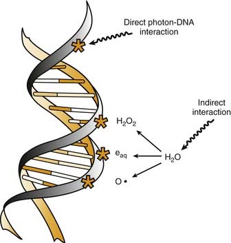
Radiation cell killing occurs when critical targets within the cell are damaged by radiation.15 A number of biologic molecules or structures are potential targets for radiation damage. According to most studies DNA is the most critical target for the biologic effects of radiation. On a molecular level, this effect requires the production of ionizations, which is why we refer to the process as ionizing radiation. The damage can occur directly when the radiation is absorbed by the DNA itself, because the atoms of the DNA become ionized and damaged. More commonly, however, it occurs indirectly through the following three modes of action15,16:

All of these species are highly reactive free radicals that, in turn, interact with the DNA and cause damage. Both ways eventually cause broken bonds in the DNA backbone, which can cause double-strand breaks, ultimately resulting in mitotic death (Fig. 77-6).

These broken bonds can result in the loss of a base or of the entire nucleotide, or in complete breaking of one or both of the strands of DNA. Single-strand breaks are easily repaired with use of the opposite strand as a template. Therefore, single-strand breaks show little relation to cell killing, although they might result in mutation if the repair is incorrect. Double-strand breaks, on the other hand, are thought to be the most important lesion in DNA produced by radiation.18 Double-strand breaks, as the name implies, results in snapping of the chromatin into two pieces. These double-strand breaks can result in mutations or, most important, in cell killing.

A growing body of experimental data suggests that radiation damage to DNA is not the only mechanism by which ionizing radiation damages cells. Other mechanisms are apoptosis, cell cycle arrest, and mitotic death. One study has suggested that apoptosis can be triggered by radiation energy deposition in cell membranes.19 It has also been reported that direct radiation damage to mitochondria can trigger apoptosis.20

Cell Cycle Arrest

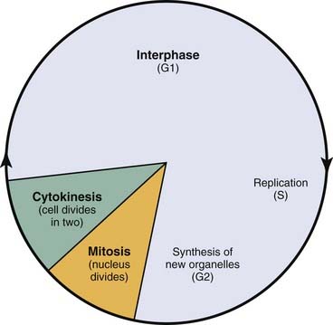
Radiation triggers signaling cascades leading to arrest, usually at the G1 and G2 checkpoints in the cell cycle.21 Cell cycle perturbations (Fig. 77-7) are seen characteristically after radiation exposure and were among the earliest observed biologic effects of radiation. Cells can show checkpoints or arrest in any phase of the cell cycle, although the best-described checkpoints with respect to radiation damage are the G1 and G2 checkpoints. Normal cells and those cancer cells that retain p53 function are blocked in the G1 phase of the cell cycle. This is a p53-mediated event.

Radiosensitivity

Our main interest in radiobiology is in figuring out ways to improve the treatment toxicity ratio. One of the main concepts is radiation sensitivity, which refers to the relative susceptibility of cells, tissues, tumors, or organisms to radiation. However, tumor regression is not solely a function of tumor cell death, but is influenced by many factors, including the amount of extracellular stroma, the propensity of the tumor cells to undergo rapid rather than delayed death, and the resorption of radiation-inactivated cells (depopulation). A frequent misconception is that tumors should be more radiosensitive than normal tissues because they proliferate more rapidly. This misconception may go back 100 years. In 1906, only 11 years after the discovery of x-rays, Bergonie and Tribondeau formulated a “law” for the relationship between cellular radiosensitivity and reproductive capacity.22 They postulated that cells that have a higher proliferation rate are more radiosensitive than slowly proliferating cells. On a purely cellular basis, it is correct that cells in mitosis at the time of irradiation are more radiosensitive than cells in other phases of the cell cycle. However, many other factors, such as tissue- and host-specific factors, have important influence. In 1906, there was no appreciation of late-occurring normal tissue complications. Nowadays, we know that many slowly proliferating or nonproliferating normal tissues, such as the kidney, are highly radiosensitive; they just express radiation injury much later than rapidly proliferating tissues. Similarly, the proliferation rate of tumors does not predict their radio-curability. For example, rapidly proliferating tumors such as glioblastoma multiforme can be highly radioresistant.

Cell Survival

Loss of reproductive integrity in long-term survival assays is important to our understanding of the response of either a tumor or a normal tissue to radiation. When cells are exposed to lethal doses of radiation, they may not die immediately or within a few hours of treatment, or even sometimes within a single division of radiation. When cells have been observed by time-lapse cinematography after irradiation, some cells survive and go on to form colonies, and some die quickly. Others go through up to several rounds of abortive cell division before finally ceasing to divide. Radiation biologists have demonstrated that it is the proportion of cells capable of forming a colony by sustained cell division that most fully predicts the effects of a dose of radiation.23 In order to eradicate or control a tumor; one must inactivate all clonogenic tumor cells. In other words, the treatment may fail if only one clonogen survives, because that cell can give rise to a regrowing tumor. In order to understand tumor control better, plots of the surviving fraction of cells as a function of the radiation dose have been performed. An example is shown in Figure 77-8.

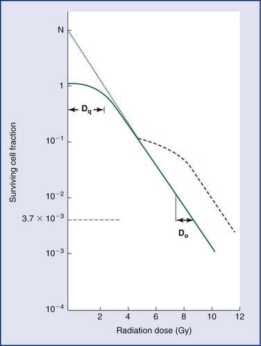
By convention, the surviving cell fraction is plotted on a logarithmic scale, and the radiation dose is plotted on a linear scale. This curve is representative of most mammalian cells. Consider the solid curve in Figure 77-8, which represents the survival data. Note that there are two distinct regions to the curve. There is an initial region for low radiation doses, where the slope of the curve is shallow. In this region, small incremental changes in the amount of radiation are not very effective at increasing the number of cells killed. This is called the shoulder region, and its width is characterized by the parameter Dq. It is the distance along the dose axis at a surviving fraction of unity between the abscissa and the point where the extrapolated linear portion of the curve is intersected. It is a measure of the ability of the cells to repair small amounts of radiation damage. At higher doses of radiation, the curve becomes a straight line on a semilog plot. Its slope is characterized by Do, which is the incremental dose change required to reduce the surviving cell fraction to 1/e of its value. The steeper the slope in this region, the smaller is the value of Do and the more radiosensitive is the cell line. When extrapolated back to a zero radiation dose, it intersects the abscissa at a value N. A curve of this type can be modeled using the following equation:

(Eq. 77-3) image

where S is the surviving fraction, D is the radiation dose, and N and Do are as indicated in Figure 77-8. In target theory, N can be thought of as the number of distinct targets in the cell that should receive one radiation “hit” before the cell is inactivated.

A number of mathematical models have been devised to attempt to describe the shape of the cell survival curves that are observed experimentally, with an initial shallower slope, an eventual bending (the “shoulder”), and a final, steeper slope. These include target models, lethal and potentially lethal damage models, and repair saturation models. Some of these have been based on simple mathematical modeling without any real attempt to model known molecular events involved in cell killing, whereas others have been based on attempts to model some of the known molecular events (e.g., chromosome breaks or DNA repair) that are involved in cell killing. All of the models can describe the shape of the survival curve to a first approximation. None does so perfectly, and none takes into account all the events and all of the possible mechanisms involved in cell death. A model that has been most influential on clinical practice is the linear quadratic model,24 because it is one of the models that best fits the behavior of cells after exposure to radiation doses within the range used in the clinic. The linear-quadratic model was devised by Keller and Rossi,25 who proposed that radiation-induced cell killing resulted from two potential events, one with a linear relation to dose (exp[−αD]), the other having a quadratic relation to dose (exp[−βD2]). This was expressed mathematically by the “alpha-beta” equation, as follows:

(Eq. 77-4) image

where S represents survival of a cell population after a dose, D (Fig. 77-9). It has been hypothesized that this equation actually represents a molecular reality.26 Double-strand breaks in the DNA are lethal lesions that are produced by either one energy deposition, an event termed (αD), or by two separate events, each involving a single strand of DNA (βD2), which then interact. This hypothesis is now thought to be unlikely because of the low probability of two tracks interacting within a single double helix.

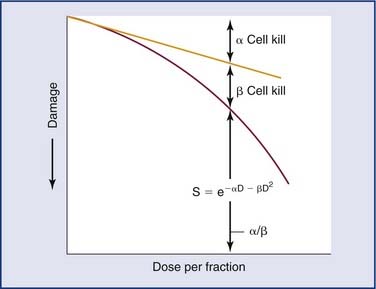
The α/β ratio, which is derived from Equation 77-4, represents a point on the survival curve at which the components of cell killing are equal to each other—that is, αD = βD2, or D = α/β. In other words, for each cell population there is a dose of radiation in which the linear (α) and quadratic (β) contributions to cell killing are equal. The α/β ratio is specific to each cellular population and reflects the sensitivity of the cell to the two types of damage. Tissues that have an early response to radiation (skin, mucosa, and tumor cells) have a high α/β ratio. In other words, their survival curves stay straight for a longer period before the bend occurs, in which there is a higher contribution of single-event or α killing. Late-responding tissues, such as spinal cord, kidney, and muscle, have survival curves that bend earlier, with resultant lower α/β ratios. These late-responding tissues have shoulders on their survival curves within the range of doses commonly used in RT. This understanding led to the concept that altered fractionation schedules could be used to treat tumor populations more effectively with respect to damage to late-responding tissues.