Neuroblastoma

Published on 06/06/2015 by admin

Filed under Pediatrics

Last modified 06/06/2015

Print this page

rate 1 star rate 2 star rate 3 star rate 4 star rate 5 star
Your rating: none, Average: 5 (1 votes)

This article have been viewed 10108 times

58 Neuroblastoma

Neuroblastoma is the most common extracranial solid tumor of childhood and accounts for 8% to 10% of all childhood cancers. The median age at diagnosis is 17 months, and the vast majority of neuroblastoma is diagnosed before age 10 years. The prognosis varies significantly and depends on age, tumor biology, and extent of disease at diagnosis. Whereas some disseminated tumors in infants may spontaneously regress, metastatic tumors diagnosed in slightly older children carry a long-term survival rate of around 40%. Although many children with neuroblastoma will need aggressive surgical resection and chemotherapy, some children can be safely observed over time. It is this dramatic heterogeneity of phenotypes that has prompted further evaluation of the implications of staging and treatment. Novel therapies are becoming increasingly important in treating refractory disease.

Etiology and Pathogenesis

Neuroblastoma is characterized as a small, round, blue-cell tumor and arises from neural crest cells. The Shimada and Joshi staging system is based on the pathologic components of neuroblastoma. The important histologic components for classification are degree of neuroblast cell differentiation, mitosis-karyorrhexis index (MKI), and amount of stromal content. This combined with age at diagnosis divides patients into two categories: favorable and unfavorable histology.

Additionally, a number of genetic aberrations are important in classification and prognosis of neuroblastoma. The DNA content of tumor cells is characterized as near-diploid or hyperdiploid, the latter of which is associated with a more favorable outcome. Examples of other genetic abnormalities associated with prognosis include MYCN amplification, deletion of the short arm of chromosome 1p, and deletion of chromosome 11q. The most important of these, MYCN amplification, is associated with rapid tumor progression and poor prognosis, even in patients with otherwise lower stage disease. Neuroblastoma is most commonly an isolated diagnosis, but it has been associated with other neurocristopathies such as Hirschsprung’s disease, congenital central hypoventilation syndrome (CCHS or Ondine’s curse), and neurofibromatosis type 1. Additionally, a very small number of patients (1%-2%) present as part of a familial neuroblastoma syndrome. In this situation, the most common genetic abnormality is a germline mutation in the anaplastic lymphoma kinase (ALK) gene (Figure 58-1).

Clinical Presentation

Neuroblastoma can arise anywhere along the sympathetic chain, and symptoms at the time of diagnosis depend on the location and extent of disease. Primary tumors most commonly occur in the abdomen (65%) and usually present as a painless abdominal mass. These patients may also present with vomiting, constipation, or symptoms of intestinal obstruction. Other common sites of primary tumors include the neck, chest, and paraspinal region. Tumors arising from the chest may be found incidentally on chest radiography, and cervical tumors may present as a Horner’s syndrome or more rarely with superior vena cava syndrome (Figure 58-2).

Approximately half of patients have metastatic disease at the time of diagnosis. Infants with metastatic disease often present with massive hepatomegaly with or without respiratory compromise. Infants may also present with bluish, subcutaneous nodules, a hallmark of the disease in this population. Other symptoms of metastatic disease in any patient include anorexia, bone pain, irritability, fever, pallor, and hypertension (most often as a result of renal vascular compression). Periorbital ecchymosis and proptosis (from bony tumor infiltrate) are also characteristic of neuroblastoma. Rarely, children are symptomatic from tumor cell catecholamine release, resulting in flushing, sweating, headache, palpitations, and hypertension.

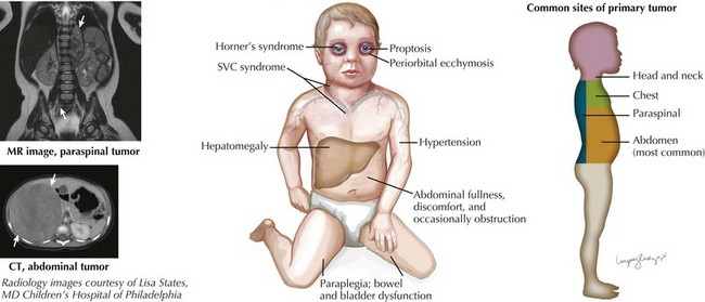
A small percentage of children (5%) present with symptoms of spinal cord compression. This oncologic emergency is associated with paraspinal neuroblastomas, and associated findings include lower extremity weakness, bowel and bladder dysfunction, back pain, and sensory loss. Neurologic function at the time of diagnosis has a strong association with long-term neurologic outcome (see Figure 58-2).

In addition, distinct paraneoplastic syndromes are associated with neuroblastoma. Secretion of vasoactive intestinal peptide by tumor cells results in intractable, watery diarrhea; hypokalemia; and poor growth. These tumors are often associated with favorable histology and good prognosis. The syndrome of opsoclonus–myoclonus is characterized by rapid, involuntary eye movements in all directions; irregular, frequent muscle jerking; and ataxia. These children usually also have a low-stage tumor with favorable biologic features. The presenting neurologic symptoms of opsoclonus–myoclonus and ataxia typically resolve after treatment; however, the majority of children have residual developmental delay.

The previously described clinical symptoms should be carefully evaluated in the physical examination. The abdominal examination should focus on the presence of a mass or hepatomegaly. Head and neck evaluation should include examination for proptosis, periorbital ecchymosis, and Horner’s syndrome (ptosis, miosis, anhidrosis, and ipsilateral facial flushing). One should perform an examination of the cervical, supraclavicular, axillary, and inguinal areas for lymphadenopathy. It is essential to complete a careful neurologic examination because subtle findings can indicate an evolving paraspinal mass. Key neurologic findings include lower extremity weakness, hyperreflexia or diminished weakness, decreased rectal sphincter tone, bowel or bladder incontinence, and paraplegia.