Neoplasms of the pineal gland
 NEOPLASMS IN THE PINEAL GLAND

 PINEAL PARENCHYMAL NEOPLASMS
 Approximately 20% of pineal parenchymal neoplasms are pineocytomas, 30% are pineoblastomas, and 50% are intermediate neoplasms.

 Patients presenting with pineal parenchymal neoplasms are mostly in their 3 rd or 4th decade.
 There is an equal incidence of pineal parenchymal neoplasms in males and females.

 Rarely, pineoblastomas are associated with bilateral retinoblastomas in children.
 Classic pineocytomas respond relatively well to surgery and focal radiotherapy.

PINEAL PARENCHYMAL NEOPLASMS


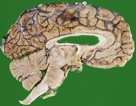
MACROSCOPIC APPEARANCES
Pineocytomas are firm gray neoplasms. Pineoblastomas are softer and sometimes gelatinous in texture and may contain areas of hemorrhage and necrosis. Although both types of neoplasm may be circumscribed, pineocytomas usually compress surrounding tissues (Fig. 39.1), while pineoblastomas tend to infiltrate the midbrain, third ventricle, or sometimes the thalamus and hypothalamus. Pineoblastomas frequently disseminate throughout the CNS along CSF pathways.
MICROSCOPIC APPEARANCES
Pineocytoma
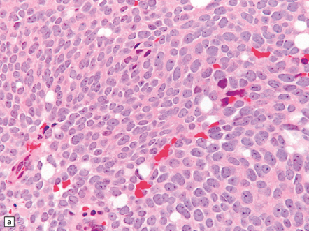
The pineocytoma consists predominantly of small monomorphic cells set in a fibrillary background (Fig. 39.2). The cells are generally arranged in sheets, though a nodular pattern may occasionally be evident. A characteristic feature is the formation of pineocytomatous rosettes, which are relatively large zones of fine, fibrillary processes surrounded by an oval arrangement of neoplastic cell nuclei (see Fig. 39.5). The neoplastic cells generally resemble the cells of normal pineal gland, with their argyrophilic processes which may have club-like terminal expansions. Their processes express NSE and synaptophysin, and at the ultrastructural level contain microtubules and a few dense core vesicles. Some cells label with antibodies to S antigen.
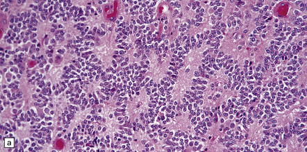
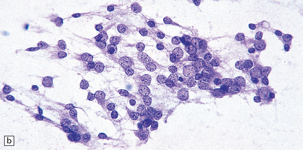
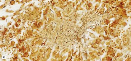
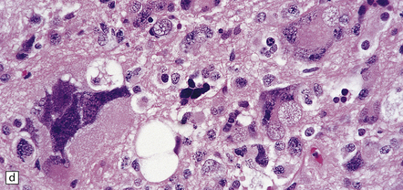
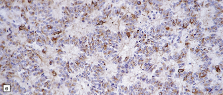
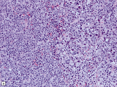
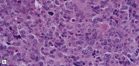
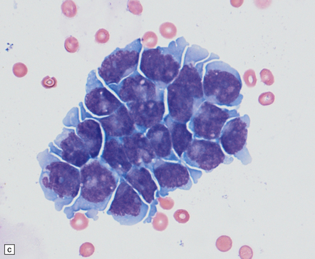
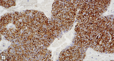
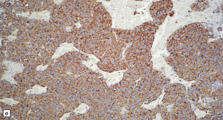
39.2 Pineocytoma.
(a) Pineocytomatous rosettes dominate this region of a typical pineocytoma. (b) In a smear preparation, the cells of this pineocytoma are characterized by uniformity, delicate cytoplasmic processes, and tiny nucleoli. (c) Silver impregnation of the neuritic processes in a pineocytomatous rosette. (d) Cytologic pleomorphism as marked as here may be seen in pineocytomas and intermediate pineal parenchymal neoplasms. In an otherwise typical pineocytoma with pineocytomatous rosettes, but without mitoses and focal increases in nuclear:cytoplasmic ratio, it is not thought to be an adverse prognostic sign. (e) Widespread immunoreactivity for neurofilament proteins is typical of pineocytomas.
Intermediate or mixed pineal parenchymal neoplasms
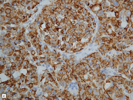
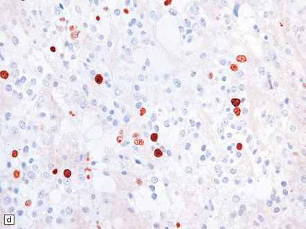
 The pineal parenchymal tumor of intermediate differentiation is characterized by a syncytial or occasionally lobular architecture (Fig. 39.3). Some of its cells may resemble those of a pineocytoma, but it lacks the well-differentiated features of this tumor, including pineocytomatous rosettes. Collections of ‘blast’ cells are also missing. Cytologic pleomorphism is variable, but usually not marked. Mitoses are readily found. Ganglion cells and giant cells are occasionally evident. Immunoreactivity for neuronal markers may be prominent. This variant sometimes metastasizes throughout the CNS. Indicators of a more favorable outcome among intermediate tumors are immunoreactivity for neurofilament proteins, and a mitotic count of less than 6/10 hpfs.
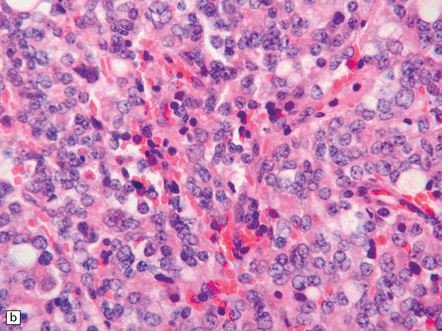

Pineoblastoma
The pineoblastoma resembles the classic (non-desmoplastic) medulloblastoma, and was previously grouped with other embryonal neoplasms of neuroepithelial lineage in the broad category of primitive neuroectodermal tumors (Chapter 38). It contains round or polyhedral cells with a high nuclear:cytoplasmic ratio, which are arranged in sheets or lobules (Fig. 39.4). Mitoses and apoptotic bodies are generally plentiful. Neuroblastic (Homer Wright) and Flexner–Wintersteiner rosettes may occasionally be found, but pineocytomatous rosettes are not a feature of the typical pineoblastoma (Fig. 39.5), being found only in the rare mixed tumor. Necrosis and hemorrhage are common, while melanin-producing cells or signs of mesenchymal differentiation, such as striated muscle or cartilage, are exceptional. Immunoreactivity for neuronal markers may be local or widespread (Fig. 39.4), and pineoblastomas may label with antibodies to retinal S antigen.