Morphea
Management strategy
Initial evaluation
The initial evaluation of patients with morphea should involve:
 Questions regarding recent development of new lesions/extension of existing lesions to determine level of activity


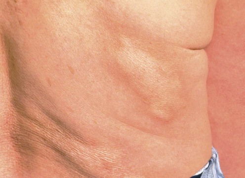
 Palpation of affected areas for depth of sclerosis


 Imaging with MRI if musculoskeletal involvement is suspected.
Active morphea lesions should be treated as the lesions are frequently symptomatic and can produce permanent cosmetic and functional sequelae. Initial evaluation should be focused on determining the extent, severity, and activity of morphea lesions laying the groundwork for rational therapeutic choices.


