Kidney Anatomy and Physiology

Published on 07/03/2015 by admin

Filed under Critical Care Medicine

Last modified 07/03/2015

Print this page

rate 1 star rate 2 star rate 3 star rate 4 star rate 5 star
Your rating: none, Average: 0 (0 votes)

This article have been viewed 6412 times

Kidney Anatomy and Physiology

Mary E. Lough

The kidneys are complex organs responsible for numerous functions and substances necessary to maintain homeostasis. The primary roles of the kidneys are to remove metabolic wastes, maintain fluid and electrolyte balance, and help achieve acid–base balance. Hormones produced by the kidneys have an important role in blood pressure control, red blood cell production, and bone metabolism. The kidneys are important in maintaining the intracellular and extracellular environment required by all cells to function effectively. When a patient experiences kidney dysfunction, some or all of the functions of the kidneys may be decreased or absent, leading to altered homeostasis.

This chapter provides an overview of the anatomy and physiologic processes of the kidneys. An understanding of normal kidney function is essential to understanding the pathophysiology, symptoms, and therapeutic management of kidney disease and failure.

Macroscopic Anatomy

The kidneys are paired organs located retroperitoneally, one on each side of the vertebral column between T12 and L3.1 The right kidney is slightly lower than the left because of the position of the liver. Each kidney is approximately 12 centimeters (cm) long, 6 cm wide, and 2.5 cm thick in the adult. Kidney size and weight varies between men and women; 125 to 170 grams in men, and 115 to 155 grams in women.1 The kidneys are protected anteriorly and posteriorly by the rib cage and by a tough fibrous capsule that encloses each kidney. Additional protection is provided by a cushion of perirenal fat and the support of the kidney fascia.

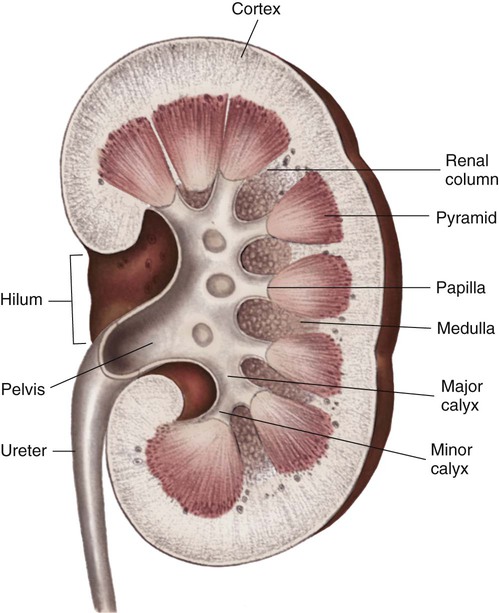
Internally, the kidneys are made up of two distinct areas: the cortex and the medulla. The kidney cortex is the outer layer and contains the glomeruli, proximal tubules, cortical portions of the loops of Henle, distal tubules, and cortical collecting ducts. The kidney cortex is about 1 cm in thickness. The kidney medulla is the inner kidney layer, made up of the pyramids, which contain the medullary portions of the loops of Henle, the vasa recta, and the medullary portions of the collecting ducts. Numerous pyramids taper and join to form a minor calyx; several minor calyces join to form a major calyx. The major calyces then join and enter the funnel-shaped kidney pelvis, a 5- to 10-mL conduit that directs urine into the ureter (Fig. 25-1).

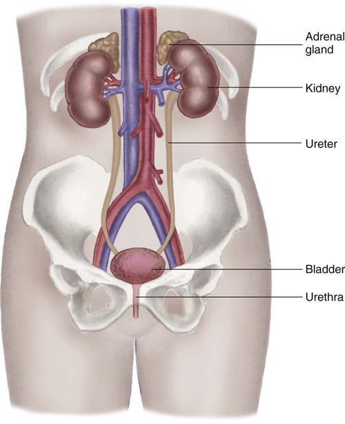
The kidney system also includes the urinary drainage system—the ureters, bladder, and urethra (Fig. 25-2). The ureters are fibromuscular tubes that exit the central part of the kidney pelvis. The ureters are 28 to 34 cm in length and enter the urinary bladder at an oblique angle. As urine is formed by the kidneys, the urine flows through the ureters by peristalsis. The peristaltic action of the ureters and the angle at which the ureters enter the bladder help prevent reflux of urine from the bladder back up into the kidneys. The bladder is a muscular sac within the pelvis and has a capacity of 280 to 500 mL. Urine leaves the bladder through the urethral orifice and is excreted from the body through the urethra. The male urethra is about 20 cm long; the female urethra is 3 to 5 cm long.

Vascular Anatomy

The kidneys are highly vascular and receive up to 20% of the cardiac output—about 1 liter to 1.2 L/min of blood flow.2 Blood enters the kidneys through the renal arteries, which branch bilaterally from the abdominal aorta. The renal artery divides into arterial branches that become progressively smaller vessels, eventually ending with the afferent arterioles. A single afferent arteriole supplies blood to each glomerulus, a tuft of capillaries that is the first structure of the nephron. The nephron is often described as the functional unit of the kidneys.1

Blood exits the glomerulus by the efferent arteriole, which connects with the peritubular capillary network, also known as the vasa recta (straight vessels), that parallel the long loops of Henle. The intricate capillary network maintains the intracapillary pressure that allows water and solutes to move between the tubules and the capillaries for urine formation and the concentration and dilution of urine. The capillaries then rejoin and form gradually enlarging venous vessels, until the blood leaves each kidney through the renal vein and returns to the general circulation by the inferior vena cava.2

Microscopic Structure and Function

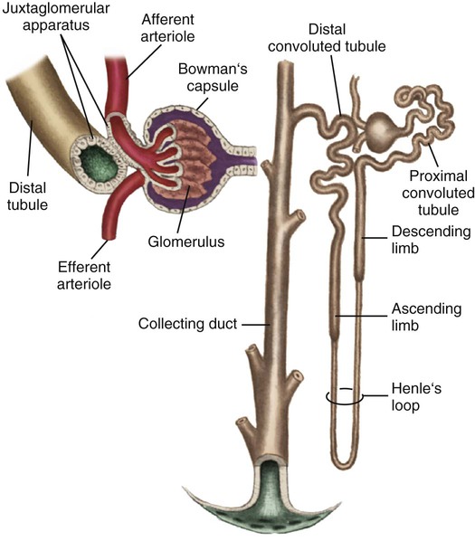
Each kidney is made up of about one million nephrons, the functional units of the kidneys. Because of the vast number of nephrons, the kidneys can continue to function even when several thousand nephrons are damaged or destroyed by disease or injury. Each nephron has the ability to perform all of the individual functions of the kidneys. The nephron is made up of several distinct structures: the glomerulus, the Bowman capsule, the proximal tubule, the loop of Henle, the distal tubule, and the collecting duct (Fig. 25-3).

Two types of nephrons make up each kidney: the cortical nephrons and the juxtamedullary nephrons. Most are cortical nephrons. These superficial cortical nephrons have glomeruli located in the outer cortex and have short loops of Henle. The midcortical nephrons are located lower in the cortex and have loops of Henle that may be short or long. Both types of cortical nephrons perform excretory and regulatory functions. The remaining nephrons are juxtamedullary nephrons with glomeruli located deep in the cortex and extending into the medullary layer of the kidney. The juxtamedullary nephrons have long loops of Henle that have an important role in the concentration and dilution of urine. The peritubular capillaries, known as the vasa recta, surround the juxtamedullary nephrons maintaining a concentration gradient to concentrate the urine.

Glomerulus

The first structure of each nephron is the glomerulus, a high-pressure capillary bed that serves as the filtering point for the blood. Positive filtration pressure in the glomerulus is achieved as a result of the high arterial pressure as the blood enters the afferent arteriole and the resistance created by the smaller efferent arteriole as the blood exits the glomerulus. As a result of the positive-pressure gradient, fluid and solutes are filtered through the glomerular capillary walls. The glomerulus has three layers: the endothelium, the basement membrane, and the epithelium. The inner endothelial layer lines the glomerulus and contains numerous pores that allow filtration of fluid and small solutes from the blood. The middle basement membrane layer also controls filtration according to the size, electrical charge, protein-binding capability, and shape of the molecules. This complex is also described as the glomerular filtration barrier (GFB). It is freely permeable to water and small or midsized molecules but large molecules such as albumin and red blood cells are prevented from entering the filtrate.3 The presence of large molecules in the urine is a signal that the glomerular membrane is damaged. The outer epithelium layer contains pores that allow the filtered blood, or filtrate, into the Bowman space.

Proximal Tubule

The proximal tubule is located in the cortex of the kidney and has a large surface area available for solute and fluid transportation. The proximal tubule resorbs (takes back) most of the filtered water and sodium and many of the solutes the body does not routinely excrete in the urine. Solutes that are usually resorbed include all of the glucose, some of the water-soluble vitamins, most phosphate and bicarbonate, and much of the potassium, chloride, and calcium that is filtered by the glomerulus. Proteins are resorbed in the proximal tubule by two specialized receptors known as megalin and cubilin that bind albumin and vitamin-binding proteins.1 Creatinine is minimally resorbed and is excreted in the urine.

In addition to its major role in resorbing water and solutes from the filtrate, the proximal tubule secretes organic anions and cations into the tubular lumen. Ammonia is produced from the metabolism of glutamine in the mitochondria of the proximal tubule cells, where ammonia (NH3) combines with hydrogen (H+) to form ammonium (NH4+), which is secreted into the proximal tubule lumen.1

Because of the large amount of solutes in the glomerular filtrate, the fluid that enters the proximal tubule is hyperosmotic. When the filtrate leaves the proximal tubule and enters the loop of Henle, it is isosmotic (equivalent to plasma) as a result of the resorption of solutes and water.

Loop of Henle

After selective resorption in the proximal tubule, the isosmotic filtrate enters the loop of Henle. The loop of Henle consists of a thin descending limb, a thin ascending limb, and a thick ascending limb. There are two types of nephrons: the cortical nephrons with short loops of Henle and the juxtamedullary nephrons with long loops of Henle. The nephrons with short loops of Henle do not have a thin ascending limb. As a result, the cortical nephrons perform excretory and regulatory functions but play only a minor role in the concentration or dilution of urine. The juxtamedullary nephrons have glomeruli that are next to (juxtaposed to) the medulla near where the cortex and medulla sections of the kidney join and contain a thin ascending limb. These nephrons with the thin ascending limbs are critical for concentrating and diluting the urine by means of the countercurrent mechanism. The thin descending limb is very permeable to water but fairly impermeable to urea, sodium, and other solutes. As a result, water (but not solute) is resorbed into the general circulation, and a more concentrated filtrate is produced. The filtrate then moves up the thin ascending limb, which is impermeable to water but allows movement of sodium, chloride, and urea back into the filtrate. The thick ascending limb is also impermeable to water but allows resorption of sodium, chloride, potassium, calcium, and bicarbonate. Because of the low water and high solute resorption in the loop of Henle, the filtrate leaves the ascending limb hypo-osmotic (more dilute than plasma).

Distal Tubule

The hypo-osmotic filtrate enters the distal tubule located in the cortex of the kidney. The first portion of the distal tubule contains the cells of the macula densa, which are specialized cells that are a component of the juxtaglomerular apparatus important in blood pressure control. The first section of the distal tubule is impermeable to water and transports solutes such as sodium, bicarbonate, calcium, and potassium. The later section of the distal tubule further regulates sodium, bicarbonate, potassium, and calcium according to hormonal influences and the acid–base and electrolyte balance needs of the body. The permeability of the late distal tubule is influenced by antidiuretic hormone (ADH). In the presence of ADH, the late distal tubule is impermeable to water but resorbs some solutes, and the filtrate remains hypo-osmotic. In the absence of ADH, the late distal tubule is more permeable to water, and the filtrate may become isosmotic.

Collecting Duct

Several distal tubules join to form a collecting duct that begins in the cortex and extends through the medulla to empty into the papilla. The final composition of the urine occurs in the collecting duct, primarily because of the transport of potassium, sodium, and water. Water permeability is determined by the absence or presence of ADH. In the absence of, or with small amounts of ADH, the urine becomes dilute, whereas larger amounts of ADH result in concentrated urine. The filtrate usually is more concentrated when it leaves the collecting duct than it was when it entered. Acidification of the urine is accomplished by the transport of bicarbonate and hydrogen in the collecting duct. Several collecting ducts then combine to form the pyramids. After the urine leaves the collecting ducts, no change in the composition of the filtrate occurs. Box 25-1 summarizes tubular resorption and secretion in the various structures of the nephron.

Nervous System Innervation

The autonomic nervous system provides the primary innervation to the kidneys and the urinary drainage system. Kidney neural innervation is derived from the celiac plexus and the sympathetic plexuses of the abdominal viscera to form the renal plexus. The renal plexus enters the kidneys along the path of the renal arteries. The inferior mesenteric plexus, the hypogastric plexus, and the pudic nerve from the sacral region serve the urinary bladder, the ureters, and the urethra.

Nervous system control in the urinary tract is reflected in the process of micturition, or the release of urine. Bladder fullness stimulates stretch receptors in the bladder wall and a portion of the urethra. Signals are carried through nerves in the sacral area and return as parasympathetic messages to contract the detrusor muscle of the bladder. With a full bladder, contractions usually are powerful enough to relax the external sphincter. Sympathetic stimulation returns the external sphincter to contraction after the urine is released. The cerebral cortex and brainstem portions of the central nervous system also exert control over the urinary bladder. The central nervous system regulates the micturition reflex, frequency, and external sphincter tone and allows conscious control over release of urine from the bladder.

Urine Formation

The nephrons are responsible for removing metabolic substances and waste products from the blood and retaining essential electrolytes and water as needed by the body. The entire blood volume of an individual is filtered by the kidneys 60 to 70 times each day, resulting in about 180 L of filtrate. The glomerular filtration rate (GFR), or the amount of filtrate formed in the nephrons, is therefore about 125 mL/min. The kidneys must reduce the 180 L of filtrate to an average of 1 to 2 L of urine per day. Although 180 L of filtrate is formed, 99% of it is resorbed, and only 1% is excreted as urine. The three processes necessary for changing the 180 L of filtrate into 1 to 2 L of urine are glomerular filtration, tubular resorption, and tubular secretion.

Glomerular Filtration

The first process in urine formation is glomerular filtration, which depends on glomerular blood flow, pressure in the Bowman space, and plasma oncotic pressure.2 Glomerular blood flow is the most important of these three factors and is maintained through an autoregulatory mechanism within the kidneys.2 The autoregulatory mechanism maintains consistent kidney blood flow and perfusion at a constant level as long as the mean arterial pressure (MAP) remains between 80 and 180 mm Hg. The afferent and efferent arterioles of the glomeruli have the ability to increase or decrease the glomerular blood flow rate through selective dilation and constriction. When the mean arterial blood pressure is decreased, the afferent arteriole dilates, and the efferent arteriole constricts to maintain a higher pressure in the glomerular capillary bed and maintain the GFR at 125 mL/min. The ability of the kidneys to autoregulate blood flow begins to fail when the mean arterial blood pressure is less than 80 mm Hg or greater than 180 mm Hg.

The second factor that influences the GFR is the pressure in the Bowman space. An increase in pressure in this space decreases filtration because the increased pressure resists the movement of solutes and water from the capillaries into the space. For example, if the tubules of the nephrons are blocked by cellular debris, backward pressure is exerted on the Bowman space, the GFR drops below 125 mL/min, and urine output decreases.

The third factor that influences GFR is plasma oncotic pressure. When the oncotic pressure in the blood is decreased (as in disease states that result in low plasma protein levels), pressure in the glomerular capillary bed is decreased. Although the mean arterial pressure in the glomerulus favors filtration, decreased amounts of fluid and solutes leave the capillaries and enter the Bowman space because the oncotic pressure gradient in the plasma that encourages movement of fluid and solutes out of the plasma is less favorable. Filtration still occurs, but it is decreased from the normal 125 mL/min, resulting in a decrease in the amount of filtrate and therefore urine.

The status of the glomerular filtration system is assessed by measuring the GFR. Creatinine is used as a measure of the GFR because it is a waste product produced at a fairly constant rate by the muscles, is freely filtered by the glomerulus, and is minimally resorbed or secreted by the tubules. Most of the creatinine produced by the body is excreted by the kidneys, making the creatinine clearance a good screening and follow-up test for estimating the GFR. Creatinine clearance usually mirrors the GFR, so that a normal creatinine clearance rate is approximately 125 mL/min. A creatinine clearance rate less than 100 mL/min reflects a GFR of less than 100 mL/min and is a signal of decreased kidney function. A creatinine clearance rate (and GFR) less than 20 mL/min results in symptoms of kidney failure.

Tubular Resorption

The second process in the formation of urine is tubular resorption—the movement of a substance from the tubular lumen (filtrate) into the peritubular capillaries (blood). Tubular resorption allows the 180 L of solutes and water filtered by the glomerulus to be taken back into the circulation, decreasing the 180 L of filtrate to 1 to 2 L of urine per day. Most tubular resorption takes place in the proximal tubule and occurs by passive and active transport processes.

Passive Transport

Passive transport of substances in the tubule depends on changes in concentration gradients and does not require energy. Diffusion and osmosis are the primary passive transport processes in the nephrons. Diffusion is the spontaneous movement of molecules or solutes from an area of higher concentration to an area of lower concentration across a semipermeable membrane (not all substances cross, particularly large molecules). For example, when water is resorbed by the tubules, the concentration of urea in the tubules is increased. Urea then diffuses across the semipermeable membrane of the tubule and re-enters the plasma to achieve balance in the concentration gradient.

Osmosis is the movement of water from an area of lower solute concentration to an area of higher solute concentration. Osmosis occurs any time the concentration of solutes on one side of a semipermeable membrane is greater than the concentration of solutes on the other side of the membrane. For example, when the concentration of sodium is greater in the peritubular capillaries than in the tubules, water passively moves from the tubules into the capillaries to balance the concentration gradient.

Active Transport

Buy Membership for Critical Care Medicine Category to continue reading. Learn more here