Gastrointestinal Tract Infections

Published on 08/02/2015 by admin

Filed under Basic Science

Last modified 08/02/2015

Print this page

rate 1 star rate 2 star rate 3 star rate 4 star rate 5 star
Your rating: none, Average: 0 (0 votes)

This article have been viewed 2144 times

Gastrointestinal Tract Infections

Objectives

1. Describe the general anatomy of the gastrointestinal tract and the relationship to transmission of infectious disease.

2. Differentiate normal flora from pathogenic organisms, and describe the relative numbers of organisms distributed throughout the gastrointestinal tract.

3. Identify nonbacterial agents of infection of the gastrointestinal tract, and name their associated diseases.

4. Describe the innate immunity as it relates to the gastrointestinal tract, including physical, chemical, and bacterial components.

5. Differentiate infections of the upper and lower gastrointestinal tract based on clinical manifestations including watery diarrhea and bloody diarrhea (dysentery).

6. Identify the major cause for antimicrobial therapy–associated diarrhea and the proper laboratory diagnostic procedure for identification, including the toxin assay.

7. Identify the most common causes for watery diarrhea, dysentery, pseudomembranous colitis, and infant botulism.

8. Describe the bacterial pathogenic mechanisms associated with gastrointestinal disease, including the presence and function of enterotoxins, attachment, and invasion mechanisms.

9. Determine the adequacy of a specimen based on collection, transport, and specimen type for the diagnosis of gastrointestinal infections.

10. Define the following media, including the organisms identified and the chemical properties associated with the selection and differentiation within the media (MAC, SMAC, EMB, HEK, XLD, SS, and Campy).

11. List the organisms and microbial products that can be detected by non-culture methods.

12. Correlate patient signs and symptoms with laboratory results for the identification of the gastrointestinal pathogen.

Anatomy

We are all connected to the external environment through our gastrointestinal (GI) tract (Figure 75-1). What we swallow enters the GI tract and passes through the esophagus into the stomach, through the small and large intestines, and finally to the anus. During passage, fluids and other components are added to this material as secretory products of individual cells and as enzymatic secretions of glands and organs, and they are removed from this material by absorption through the gut epithelium.

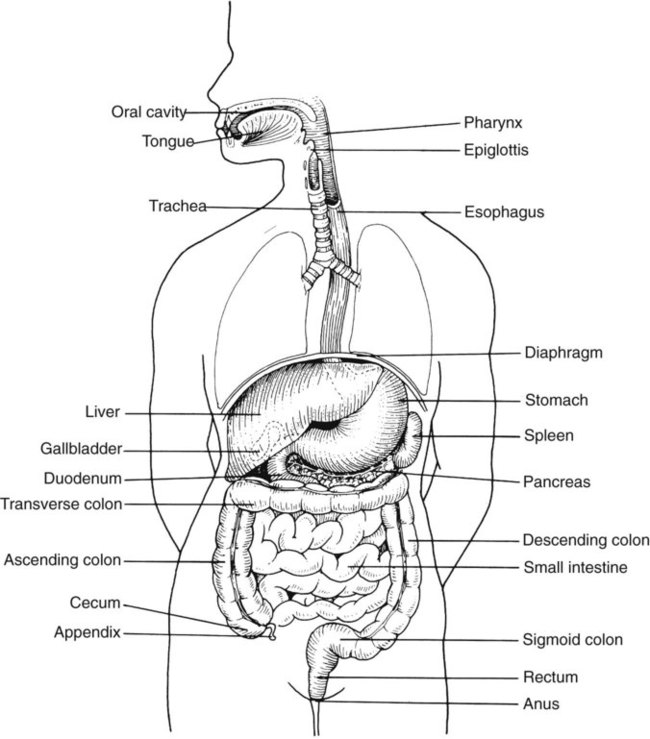
The major components of the tract are listed in Box 75-1. The nature of the epithelial cells lining the GI tract varies with each portion. The lining of the GI tract is called the mucosa. Because of the differing nature of the mucosal surfaces of various segments of the bowel, specific infectious disease processes tend to occur in each segment.

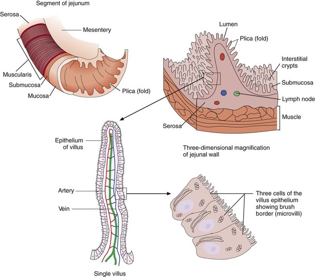
The wall of the small intestine has folds that have millions of tiny, hairlike projections called villi. Each villus contains an arteriole, venule, and lymph vessel (Figure 75-2). The function of villi is to absorb fluids and nutrients from the intestinal contents. Epithelial cells lining the surface of villi have a surface resembling a fine brush, referred to as a brush border. The brush border is formed by nearly 2000 microvilli per epithelial cell. Intestinal digestive enzymes are produced in brush border cells toward the top of the villi. Villi and microvilli help make the small intestine the primary site of digestion and absorption by significantly increasing the surface area; more than 90% of physiologic net fluid absorption occurs here. Mucus-secreting goblet cells are found in large numbers of villi and intestinal crypts.

Similar to the small intestine, the large intestine is composed of several segments (see Box 75-1). The wall of the large intestine consists of columnar epithelial cells, many of which are mucus-producing goblet cells. In contrast to the small intestine, there are no villous projections into the lumen. The remaining excess fluid within the GI tract is resorbed through the cells lining the large intestine before waste is finally discharged through the rectum.

In addition to the previously discussed components of the GI tract, numerous other organs and structures are either located in the main digestive organs or open into them. These accessory organs and structures include the salivary glands, tongue, teeth, liver, gallbladder, and pancreas. Except for the teeth and salivary glands, these organs are illustrated in Figure 75-1.

Resident Microbial Flora

The GI tract contains vast, diverse normal flora. Although the acidity of the stomach prevents any significant colonization in a normal host under most circumstances, many species can survive passage through the stomach to become resident within the lower intestinal tract. Normally, the upper small intestine contains only sparse flora (bacteria, primarily streptococci; lactobacilli; and yeasts; 101 to 103/mL), but in the distal ileum, counts are about 106 to 107/mL, with Enterobacteriaceae and Bacteroides spp. predominantly present.

Infants usually are colonized by normal human epithelial flora, such as staphylococci, Corynebacterium spp., and other gram-positive organisms (bifidobacteria, clostridia, lactobacilli, streptococci), within a few hours of birth. Over time, the content of the intestinal flora changes. The normal flora of the adult large bowel (colon) is established relatively early in life and consists predominantly of anaerobic species, including Bacteroides, Clostridium, Peptostreptococcus, Bifidobacterium, and Eubacterium.

Aerobes, including Escherichia coli, other Enterobacteriaceae, enterococci, and streptococci, are outnumbered by anaerobes 1000 : 1. The number of bacteria per gram of stool within the bowel lumen increases steadily as material approaches the sigmoid colon (the last segment). Eighty percent of the dry weight of feces from a healthy human consists of bacteria, which can be present in numbers as high as 1011 to 1012 colony-forming units (CFU)/g of stool.

Gastroenteritis

Worldwide, diarrheal diseases are the second leading cause of death; about 48 million enteric infections occur each year. These infections cause significant morbidity and death, particularly in elderly people and children younger than 5 years of age. It has been estimated that 4 million to 6 million children die each year of diarrheal diseases, particularly in developing countries in Asia and Africa. Even in developed countries, significant morbidity occurs as a result of diarrheal illness. Although acute diarrheal syndromes are usually self-limited, some patients with infectious diarrhea require diagnostic studies and treatment.

Pathogenesis

Similar to the pathogenesis of urinary tract infections, the host and the invading microorganism possess key features that determine whether an enteric pathogen is able to cause microbial diarrhea.

Host Factors

The human host has numerous defenses that normally prevent or control disease produced by enteric pathogens. For example, the acidity of the stomach effectively restricts the number and types of organisms that enter the lower GI tract. Normal peristalsis helps move organisms toward the rectum, interfering with their ability to adhere to the mucosa. The mucous layer coating the epithelium entraps microorganisms and helps propel them through the gut. The normal flora prevents colonization by potential pathogens.

Mucous membranes line the GI tract, as well as the respiratory and urogenital tracts. These membranes are exposed to the external environment in the form of food, water, and air. These membranes contain multiple cell types; some are secreting or absorbing cells that perform physiologic functions of the membrane, while others serve as protective barriers. For example, sets of specialized cells called follicles are part of the mucous membrane lining the GI tract and serve a protective function. Collections of follicles are called Peyer’s patches. Follicles contain M cells, macrophages, and B and T cells. As a result of the collective action of the follicle components following uptake and processing of the bacteria or antigens, secretory immunoglobulin A (sIgA) is released. Phagocytic cells and sIgA within the gut help destroy etiologic agents of disease, as do eosinophils, which are particularly active against parasites. Follicles and Peyer’s patches are found in the small and large intestines.

Other factors that determine the progression and potential invasion by pathogenic organisms include the host’s personal hygiene and age. An initial step in the pathogenesis of enteric infections is ingestion of the pathogen. The majority of enteric pathogens, including bacteria, viruses, and parasites, are transmitted by the fecal-oral route. Enteric infections can be spread by contamination of food products or drinking water and then subsequent ingestion. The age of the host also plays a role in whether disease is established. For example, diarrheal infections caused by rotavirus or enteropathogenic Escherichia coli tend to affect young children.

Finally, the normal intestinal flora is an important factor in the host protection from the introduction of a potentially harmful microorganism. Whenever a reduction in normal flora occurs as a result of antibiotic treatment or some host factor, resistance to GI infection is significantly reduced. The most common example of the protective effect of normal flora is the development of the syndrome pseudomembranous colitis (PMC). This inflammatory disease of the large bowel is caused by the toxins of the anaerobic organism Clostridium difficile and occasionally other clostridia and perhaps even Staphylococcus aureus. The inflammatory disease seldom occurs except following antimicrobial or antimetabolite treatment that has altered the normal flora. Almost every antimicrobial agent and several cancer agents have been associated with the development of PMC. C. difficile, usually acquired from the hospital environment, is suppressed by normal flora. When normal flora is reduced, C. difficile is able to multiply and produce its toxins. This syndrome is also known as antibiotic-associated colitis. Other microorganisms that may gain a foothold when released from selective pressure of normal flora include Candida spp., staphylococci, Pseudomonas spp., and various Enterobacteriaceae.

Microbial Factors

The ability of an organism to cause GI infection depends not only on the susceptibility of the human host to the invading organism but also on the organism’s virulence traits. To cause GI infection, a microorganism must possess one or more factors that allow it to overcome host defenses or it must enter the host at a time when one or more of the innate defense systems are inactive. For example, certain stool pathogens are able to survive gastric acidity only if the acidity has been reduced by bicarbonate, other buffers, or by medications for ulcers (e.g., cimetidine, ranitidine, H2 blockers). Pathogens ingested with milk have a better chance of survival, because milk neutralizes stomach acidity. Organisms such as Mycobacterium tuberculosis, Shigella, E. coli O157:H7, and C. difficile (a spore-forming Clostridium spp.) are able to withstand exposure to gastric acids and thus require much smaller infectious doses than do acid-sensitive organisms such as Salmonella.

Primary Pathogenic Mechanisms.

Because the normal adult GI tract receives up to 8 L of ingested fluid daily, plus the secretions of the various glands that contribute to digestion (salivary glands, pancreas, gallbladder, stomach), of which all but a small amount must be resorbed, any disruption of the normal flow or reabsorption of fluid will profoundly affect the host. Depending on how they interact with the human host, enteric pathogens may cause disease in one or more of the following three ways:

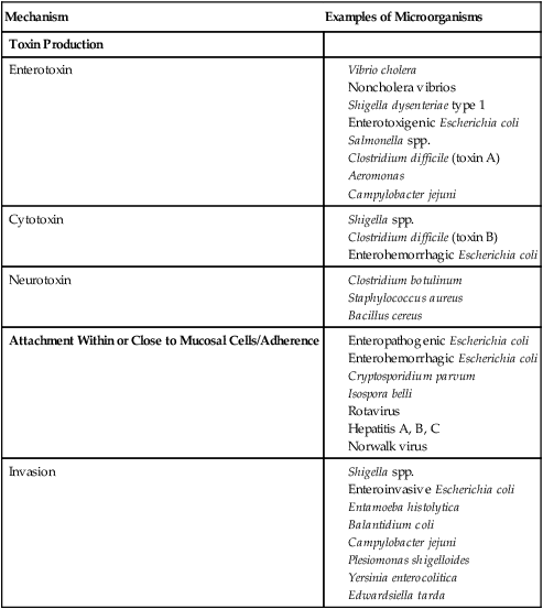
Examples of microorganisms for each of these pathogenic mechanisms are listed in Table 75-1.

Cytotoxin

Neurotoxin

Attachment Within or Close to Mucosal Cells/Adherence

Invasion

image

Toxins

Enterotoxins.

Enterotoxins alter the metabolic activity of intestinal epithelial cells, resulting in an outpouring of electrolytes and fluid into the lumen. They act primarily in the jejunum and upper ileum, where most fluid transport takes place. The stool of patients with enterotoxic diarrheal disease involving the small bowel is profuse and watery, and blood or polymorphonuclear neutrophils are not prominent features.

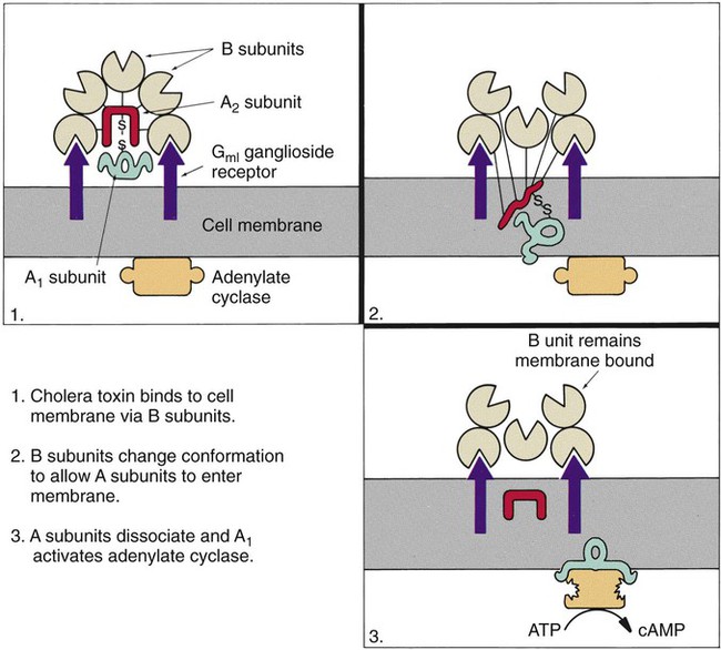
The classic example of an enterotoxin is that of Vibrio cholerae (Figure 75-3). This toxin consists of two subunits, A and B. The A subunit is composed of one molecule of A1, the toxic moiety, and one molecule of A2, which binds an A1 subunit to five B subunits. The B subunits bind the toxin to a receptor (a ganglioside, an acidic glycolipid) on the intestinal cell membrane. Once bound, the toxin acts on adenylate cyclase enzyme, which catalyzes the transformation of adenosine triphosphate (ATP) to cyclic adenosine monophosphate (cAMP). Increased levels of cAMP stimulate the cell to actively secrete ions into the intestinal lumen. To maintain osmotic stabilization, the cells then secrete fluid into the lumen. The fluid is drawn from the intravascular fluid store of the body. Patients therefore can become dehydrated and hypotensive rapidly. V. cholerae inhabits sea and stagnant water and is spread in contaminated water. The organisms have been isolated from coastal waters of several states, and sporadic cases of cholera occur in the United States. Additional information about V. cholerae is provided in Chapter 26.

Other organisms also produce a cholera-like enterotoxin. A group of vibrios similar to V. cholerae but serologically different, known as the noncholera vibrios, produce disease clinically identical to cholera, effected by a very similar toxin. The heat-labile toxin (LT) elaborated by certain strains of E. coli, called enterotoxigenic E. coli (ETEC), is similar to cholera toxin, sharing cross-reactive antigenic determinants. The enterotoxins of some Salmonella spp. (including S. enterica subsp. arizonae), Vibrio parahaemolyticus, the Campylobacter jejuni group, Clostridium perfringens, Clostridium difficile, Bacillus cereus, Aeromonas, Shigella dysenteriae, and many other Enterobacteriaceae also cause positive reactions in at least one of the tests for enterotoxin (discussed later). The exact contribution of these enterotoxins to the pathogenicity of most stool pathogens remains to be elucidated.

Certain strains of E. coli, in addition to producing a heat-labile toxin (LT) similar to cholera toxin, also produce a heat-stable toxin (ST) with other properties. Although ST also promotes fluid secretion into the intestinal lumen, its effect is mediated by activation of guanylate cyclase, resulting in increased levels of cyclic guanylate monophosphate (GMP), which yields the same net effect as increased cAMP. Tests for ST include enzyme-linked immunosorbent assay (ELISA), immunodiffusion and cell culture. Molecular techniques, including the use of DNA probes as well as several amplification assays, have been used to identify ETEC directly in clinical samples or isolated bacterial colonies.

Several tests are available for the detection of enterotoxin. Immunodiffusion, ELISA, and latex agglutination tests are all available to identify specific toxins. Molecular probes and amplification assays for toxin detection are also available, primarily for research use.

Cytotoxins.

Cytotoxins, which constitute the second category of toxins, disrupt the structure of individual intestinal epithelial cells. When destroyed, these cells slough from the surface of the mucosa, leaving it raw and unprotected. The secretory or absorptive functions of the cells are no longer performed. The damaged tissue evokes a strong inflammatory response from the host, further inflicting tissue damage. Numerous polymorphonuclear neutrophils and blood are often seen in the stool, and pain, cramps, and tenesmus (painful straining during a bowel movement) are common symptoms. The term dysentery refers to this destructive disease of the mucosa, almost exclusively occurring in the colon. Cytotoxin has not yet been shown to be the sole virulence factor for any etiologic agent of GI disease, because most agents produce a cytotoxin in conjunction with other factors.

E. coli strains seem to possess virulence mechanisms of many types. Some strains produce a cytotoxin capable of destroying epithelial cells and blood cells. Certain strains produce a cytotoxin that affects Vero cells (African green monkey kidney cells) and resemble the cytotoxin produced by Shigella dysenteriae (Shiga toxin); such strains of E. coli are associated with hemorrhagic colitis and the sequelae following infection of hemolytic-uremic syndrome (HUS) and thrombotic thrombocytopenia purpura (TTP). These strains of E. coli are referred to as enterohemorrhagic E. coli (EHEC), also referred to as serotoxigenic or STET/VTEC. See Chapter 20 for more information related to toxigenic E. coli. Table 75-2 summarizes the key pathogenic features of the primary groups of diarrheogenic E. coli.

TABLE 75-2

Overview of the Primary Groups of E. coli That Cause Diarrhea in Humans

Type Primary Mode of Pathogenesis Other Comments
Enterotoxigenic (ETEC) Produces heat-labile (LT) or heat stable (ST) enterotoxins; genes of both toxins reside on a plasmid; LTs are closely related in structure and function to cholera toxin; STs result in net intestinal fluid secretion by stimulating guanylate cyclase Common cause of traveler’s diarrhea; infects all ages
Enteroaggregative (EAEC) Binds to small intestine cells via fimbriae encoded by a large molecular weight plasmid, forming small clumps of bacteria on the cell surface; other plasmid-borne virulence factors include structured pilin, a heat-stable enterotoxin, novel anti-aggregative protein, and a heat-labile enterotoxin, all believed to be the cause of the associated diarrhea Infects primarily young children
Enteroinvasive (EIEC) Pathogenesis has yet to be totally elucidated; studies suggest that mechanisms by which diarrhea results are virtually identical to those of Shigella spp. Very difficult to distinguish from Shigella spp. and other E. coli strains
Enteropathogenic (EPEC) Initially attaches in the colon and small intestine and then becomes intimately adhered to intestinal epithelial cells, subsequently causing the loss of enterocyte microvilli (effacement); genes for attachment/effacement reside in a cluster on the bacterial chromosome (i.e., pathogenicity island) Diarrhea in infants, particularly in large urban hospitals
Enterohemorrhagic (EHEC) OR Attaches to and effaces gut epithelial cells in a similar manner as EPEC; in addition, EHEC elaborates shiga toxins Although many outbreaks are caused by E. coli O157:H7, other serotypes have been implicated in outbreaks and sporadic cases
Gene recombination among strains makes classification difficult
Enterohemorrhagic (EHEC); or serotoxigenic (STEC); verotoxigenic (VTEC) (newest, terminology) Produce one or more shiga toxins referred to as verocytotoxins. Attaches to and effaces gut epithelial cells in a similar manner as EPEC 0157 STEC serotypes; contains most common serotypes 0157 : H7 and nonmotile 0157 : NM. There are more than 150 non-0157 serotypes that have been isolated from patients with diarrhea or hemolytic uremic syndrome

C. difficile produces a cytotoxin, the presence of which is a most useful marker for diagnosis of PMC. S. dysenteriae, Staphylococcus aureus, C. perfringens, and V. parahaemolyticus produce cytotoxins that contribute to the pathogenesis of diarrhea, although they may not be essential for initiation of disease. Other vibrios, Aeromonas hydrophila (a relatively newly described agent of GI disease), and Campylobacter jejuni, the most common cause of GI disease in many areas of the United States, have been shown to produce cytotoxins. The role that these toxins play in the pathogenesis of the disease syndromes is not yet completely delineated.

Neurotoxins.

Food poisoning, or intoxication, may occur as a result of ingesting toxins produced by microorganisms. The microorganisms usually produce their toxins in foodstuffs before they are ingested; thus, the patient ingests preformed toxin. Strictly speaking, these syndromes are not GI infections but rather intoxications; because they are acquired by ingestion of microorganisms or their products, they are considered in this chapter. Particularly in staphylococcal food poisoning and botulism, the causative organisms may not be present in the patient’s bowel.

Bacterial agents of food poisoning that produce neurotoxins include Staphylococcus aureus and Bacillus cereus. Toxins produced by these organisms cause vomiting, independent of other actions on the gut mucosa. Staphylococcal food poisoning is one of the most frequently reported categories of food-borne disease. The organisms grow in warm food, primarily meat or dairy products, and produce the toxin. Onset of disease is usually within 2 to 6 hours of ingestion. B. cereus produces two toxins, one of which is preformed, called the emetic toxin, because it produces vomiting. The second type, probably involving several enterotoxins, causes diarrhea. Often acquired from eating rice, B. cereus has also been associated with cooked meat, poultry, vegetables, and desserts.

Perhaps the most common cause of food poisoning is from type A Clostridium perfringens, which produces toxin in the host after ingestion. As a result, a relatively mild, self-limited (usually 24-hour) gastroenteritis occurs, often in outbreaks in hospitals. Meats and gravies are typical foods associated with this type of food poisoning.

One of the most potent neurotoxins is produced by the anaerobic organism Clostridium botulinum. This toxin prevents the release of the neurotransmitter acetylcholine at the cholinergic nerve junctions, causing flaccid paralysis. The toxin acts primarily on the peripheral nerves but also on the autonomic nervous system. Patients exhibit descending symmetric paralysis and ultimately die of respiratory paralysis unless they are mechanically ventilated. In most cases, adult patients who develop botulism have ingested the preformed toxin in food (home-canned tomato products and canned, cream-based foods are often implicated), and the disease is considered intoxication, although C. botulinum has been recovered from the stools of many adult patients. A relatively recently recognized syndrome, infant botulism, is a true GI infection. In adults, the normal flora probably prevents colonization by C. botulinum, whereas the organism is able to multiply and produce toxin in the infant bowel. Infant botulism is not an infrequent condition; babies acquire the organism by ingestion, although the source of the bacterium is not always clear. Because an association has been found with honey and corn syrup, infants younger than 9 months of age should not be fed honey. The effect of the toxin is the same, whether ingested in food or produced by growing organisms within the bowel.