CHAPTER 48 Elbow
SKIN AND SOFT TISSUE
SKIN
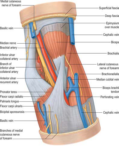
Cutaneous vascular supply
The skin overlying the cubital fossa receives its blood supply on its anterolateral side by small perforating vessels from the radial collateral artery, together with some small musculocutaneous perforators from the radial recurrent artery which traverse brachioradialis (Fig. 45.4). On the anteromedial side, skin is supplied by small branches from the anastomosis between the inferior ulnar collateral artery and the anterior ulnar recurrent artery and by small branches from the brachial artery.
Cutaneous innervation
The skin of the cubital fossa and the epicondylar regions is innervated by the medial and lateral cutaneous nerves of the forearm (Fig. 45.15). A small proximal area on the lateral aspect is supplied by the distal part of the lower lateral cutaneous nerve of the arm. The skin over the olecranon region is innervated by the distal branches of the posterior cutaneous nerve of the arm, together with proximal branches of the posterior cutaneous nerve of the forearm.
SOFT TISSUE: CUBITAL FOSSA
The cubital fossa forms a triangular depression in the middle of the upper part of the anterior aspect of the forearm (Fig. 48.1). The superior border of the fossa is an imaginary line, which joins the two epicondyles of the humerus. The fleshy elevation which constitutes its medial border is formed by the lateral margin of pronator teres and the elevation which forms the lateral border is the medial edge of brachioradialis. The roof of the fossa is formed by the deep fascia of the forearm, reinforced by the bicipital aponeurosis on the medial aspect. The median cubital vein lies on this deep fascia crossed superficially (or sometimes deeply) by the medial cutaneous nerve of the forearm. Brachialis and supinator form the floor of the fossa.
JOINTS
ELBOW JOINT
The elbow joint is a synovial joint. Its complexity is increased by continuity with the superior radio-ulnar joint. It includes two articulations (Fig. 48.2). These are the humero-ulnar, between the trochlea of the humerus and the ulnar trochlear notch, and the humero-radial, between the capitulum of the humerus and the radial head.
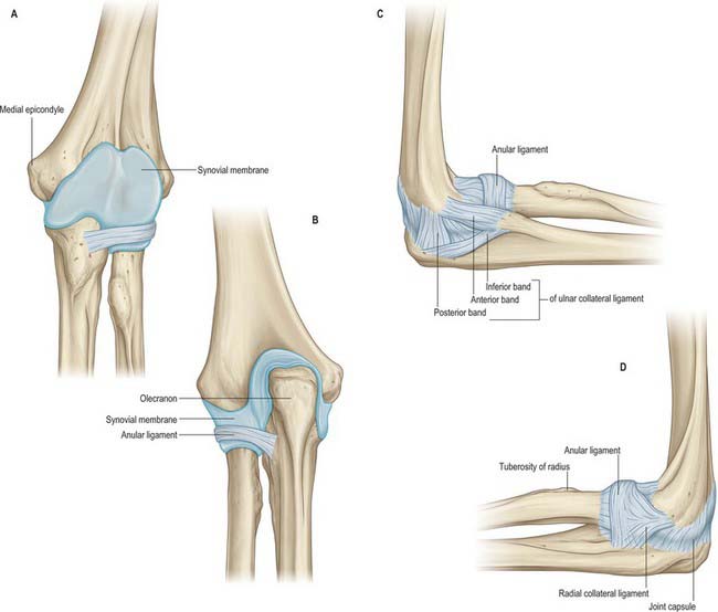
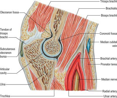
The fibrous capsule (Fig. 48.2, Fig. 48.3, 48.4) is broad and thin anteriorly. It is attached proximally to the front of the medial epicondyle and humerus above the coronoid and radial fossae, and distally to the edge of the ulnar coronoid process and anular ligament, and is continuous at its sides with the ulnar and radial collateral ligaments. Anteriorly it receives numerous fibres from brachialis. Posteriorly the capsule is thin and attached to the humerus behind its capitulum and near its lateral trochlear margin, to all but the lower part of the edge of the olecranon fossa, and to the back of the medial epicondyle. Inferomedially it reaches the superior and lateral margins of the olecranon and is laterally continuous with the superior radio-ulnar capsule deep to the anular ligament. It is related posteriorly to the tendon of triceps and to anconeus.
The humero-ulnar and humeroradial articulations have ulnar and radial collateral ligaments.
This is a triangular band, consisting of thick anterior, posterior and inferior parts united by a thin region (Fig. 48.2C). The strongest and stiffest anterior part is attached by its apex to the front of the medial epicondyle and by its broad distal base to a proximal tubercle on the medial coronoid margin. The posterior part, also triangular, is attached low on the back of the medial epicondyle and to the medial margin of the olecranon. Between these two bands intermediate fibres descend from the medial epicondyle to an inferior, oblique band, often weak, between the olecranon and coronoid processes. This converts a depression on the medial margin of the trochlear notch into a foramen, through which the intracapsular fat pad is continuous with extracapsular fat medial to the joint. The anterior band is taut throughout most of the range of flexion, while the posterior band becomes taut between half and full flexion.
This is attached low on the lateral epicondyle and to the anular ligament (Fig. 48.2D). Some of its posterior fibres cross the ligament to the proximal end of the supinator crest of the ulna. It intimately blends with attachments of supinator and extensor carpi radialis brevis. It is taut throughout most of the range of flexion.
The synovial membrane (Figs 48.2–48.4) extends from the humeral articular margins, lines the coronoid, radial and olecranon fossae, the flat medial trochlear surface, the deep surface of the capsule and the lower part of the anular ligament. Projecting between the radius and ulna from behind is a crescentic synovial fold, which partly divides the joint into humero-radial and humero-ulnar parts. Irregularly triangular, it contains extrasynovial fat (Fig. 48.5). Between the capsule and synovial membrane are three other pads of fat: the largest, at the olecranon fossa, is pressed into the fossa by triceps during flexion; the other two, at the coronoid and radial fossae, are pressed in by brachialis during extension. They are all slightly displaced in contrary movements. Smaller synovial-covered tags of fat project into the joint near constrictions flanking the trochlear notch, and cover small non-articular areas of bone.


