• Lymphoma: Bulky submucosal mass without obstruction
(Left) Spot film from from an upper GI series shows an ulcerated mass arising from the 2nd portion of the duodenum. There is a persistent pooling of barium within the lesion after the remainder of the duodenum has cleared.
(Right) Axial CECT in the same patient reveals a high-attenuation mass within the wall of the 2nd duodenum. A metastatic tumor was confirmed at surgery with the same histology as the primary colon cancer.
(Left) Small bowel follow-through in a liver transplant recipient, who presented with upper gastrointestinal pain and bleeding, shows a large amorphous collection of barium apparently arising from, and in continuity with, the distal duodenum. There is no evidence of bowel obstruction.
(Right) Axial CECT in the same patient shows a large soft tissue density mass arising from the distal duodenum. This is a good example of aneurysmal dilation of the bowel lumen caused by lymphoma.
TERMINOLOGY
Definitions
• Involvement of duodenum with malignant lymphoma or metastatic disease
IMAGING
General Features
• Best diagnostic clue
Metastases: Bull’s-eye or “target” lesion; submucosal or polypoid mass
Lymphoma: Bulky submucosal mass without obstruction of lumen
• Location
Submucosal lesion in any portion of duodenum
• Size
1-5 cm
• Morphology
Lymphoma: Smooth submucosal, often bulky mass
Fluoroscopic Findings
• Upper GI
Metastases: “Target” or bull’s-eye lesion with rounded submucosal mass; luminal obstruction and ulceration are common
–
Buy Membership for Radiology Category to continue reading. Learn more here


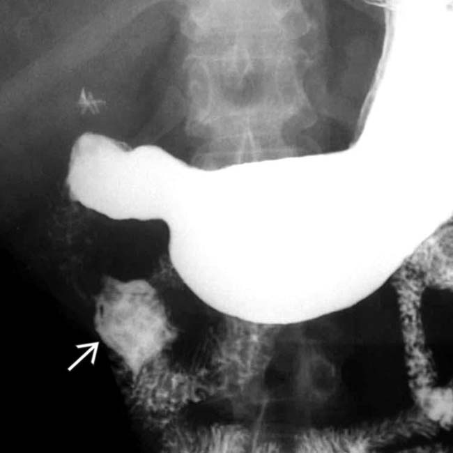
 arising from the 2nd portion of the duodenum. There is a persistent pooling of barium within the lesion after the remainder of the duodenum has cleared.
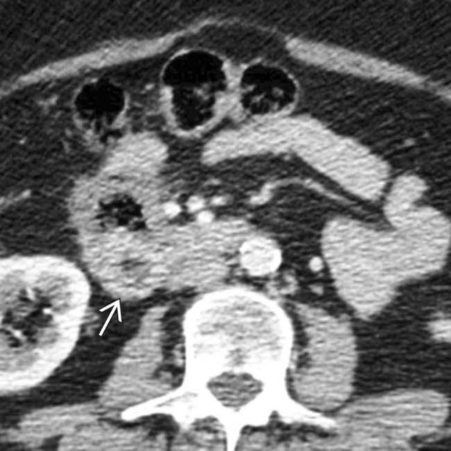
 within the wall of the 2nd duodenum. A metastatic tumor was confirmed at surgery with the same histology as the primary colon cancer.
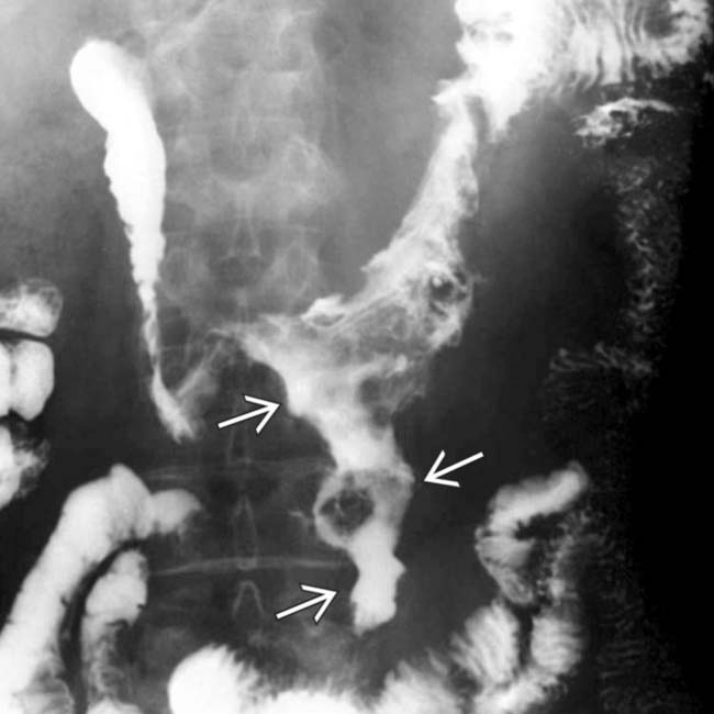
 apparently arising from, and in continuity with, the distal duodenum. There is no evidence of bowel obstruction.
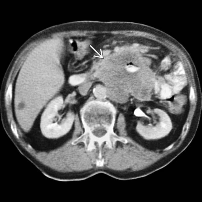
 arising from the distal duodenum. This is a good example of aneurysmal dilation of the bowel lumen caused by lymphoma.


