Definitions and Classification of Hemolytic Anemias

Published on 25/03/2015 by admin

Filed under Pediatrics

Last modified 25/03/2015

Print this page

rate 1 star rate 2 star rate 3 star rate 4 star rate 5 star
Your rating: none, Average: 0 (0 votes)

This article have been viewed 1083 times

Chapter 451 Definitions and Classification of Hemolytic Anemias

Hemolysis is defined as the premature destruction of red blood cells (RBCs). Anemia results when the rate of destruction exceeds the capacity of the marrow to produce RBCs. Normal RBC survival time is 110-120 days (half-life, 55-60 days), and approximately 0.85% of the most senescent RBCs are removed and replaced each day. During hemolysis, RBC survival is shortened, the RBC count falls, erythropoietin is increased, and the stimulation of marrow activity results in heightened RBC production, reflected in an increased percentage of reticulocytes in the blood. Thus, hemolysis should be suspected as a cause of anemia if an elevated reticulocyte count is present. The reticulocyte count also may be elevated as a response to acute blood loss or for a short period after replacement therapy for iron, vitamin B12, or folate deficiency. The marrow can increase its output 2- to 3-fold acutely, with a maximum of 6- to 8-fold in long-standing hemolysis. The reticulocyte percentage can be corrected to measure the magnitude of marrow production in response to hemolysis as follows:

image

where µ is a maturation factor of 1-3 related to the severity of the anemia (see imageFig. 451-1 on the Nelson Textbook of Pediatrics website at www.expertconsult.com). The normal reticulocyte index is 1.0; therefore, the index measures the fold increase in erythropoiesis (e.g., 2-fold, 3-fold).

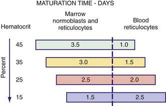
As anemia becomes more severe, the erythropoietin concentration increases and reticulocytes are released from the marrow earlier; they are identifiable as reticulocytes in the blood that last for >1 day. Because the reticulocyte index is essentially a measure of RBC production per day, the maturation factor, µ, provides this correction (see Fig. 451-1

Buy Membership for Pediatrics Category to continue reading. Learn more here