Clinical Laboratory Studies

Published on 12/06/2015 by admin

Filed under Pulmolory and Respiratory

Last modified 12/06/2015

Print this page

rate 1 star rate 2 star rate 3 star rate 4 star rate 5 star
Your rating: none, Average: 0 (0 votes)

This article have been viewed 4963 times

Clinical Laboratory Studies

Nadine A. Fydryszewski and Elaine M. Keohane

Learning Objectives

After reading this chapter, you will be able to:

1. Explain the three phases of laboratory testing.

2. Describe the composition of blood.

3. Explain the importance of specimen integrity and effects on laboratory test results.

4. Define laboratory test sensitivity, specificity, and positive and negative predictive value.

5. Discuss the meaning of the term reference range.

6. Describe the clinical applications and general clinical significance of increases and decreases for each component of the complete blood count and for the reticulocyte count and erythrocyte sedimentation rate.

7. Define leukocytosis, leukopenia, relative and absolute count, neutrophilia, neutropenia, lymphocytosis, lymphopenia, monocytosis, eosinophilia, basophilia, leukemia, anemia, and hemostasis.

8. Describe the body’s response to anemia and the potential effects on the body of uncompensated anemia.

9. Differentiate primary, secondary, and relative polycythemia and describe the adverse effects of polycythemia on the body.

10. Describe the clinical applications of the activated partial thromboplastin time, prothrombin time, and platelet count in assessing hemostasis.

11. Explain the clinical application and significance of the quantitative D-dimer assay.

12. Describe the clinical applications and general clinical significance of increases and decreases in electrolyte concentrations, glucose levels, blood urea nitrogen and creatinine.

13. Discuss the importance of renal and hepatic panel tests as related to the management of patients with respiratory disorders.

14. Relate lipid panel measures to the risk for atherosclerosis and heart disease.

15. Identify the current cardiac biomarkers used to help identify acute coronary syndrome and congestive heat failure.

16. Describe the pre-analytical phase of testing in clinical microbiology.

17. Discuss the common methods of examination of microbiology specimens (e.g., Gram stain, acid-fast stains).

18. Explain the purpose of a microbiology culture and antimicrobial sensitivity test.

19. Describe the collection and transport protocols for pulmonary secretions.

20. Discuss the importance of macroscopic and microscopic examination of sputum.

21. List the microscopic criteria used to assess the quality of a sputum sample.

22. Explain the significance of sputum eosinophilia.

23. Describe the indications and method of performing a bronchoalveolar lavage.

24. Describe the macroscopic, microscopic, and chemical significance of pleural fluid examination.

25. Explain the purpose of histologic and cytologic examinations.

26. List the malignant tumors responsible for producing most primary lung cancers.

27. List the types of pulmonary samples that can be examined cytologically.

28. Discuss the general concept of skin testing and two methods to screen for tuberculosis.

29. Given a variety of patient presentations, identify the common laboratory tests helpful in assessing the problem.

Clinical Laboratory Overview

The practice of modern medicine would be impossible without the tests performed by medical laboratory scientists in the clinical laboratory. Laboratory scientists analyze body fluids and other medical specimens, providing laboratory data and vital information to other members of the health care team. Clinical laboratory tests are used to diagnose, treat, monitor, and prevent disease.

The clinical laboratory includes several specialized disciplines: microbiology, hematology, immunology, transfusion medicine, clinical chemistry, and molecular diagnostics. Most hospitalized patients with respiratory disease undergo many laboratory tests, and it is important for respiratory therapists (RTs) to have a basic understanding of the commonly ordered tests. This chapter provides key information related to the pre-analytical, analytical, and post-analytical phases of laboratory testing. Although the emphasis is on laboratory tests and data related to patients with respiratory disease, most of the concepts described here are applicable to any patient.

Phases of Laboratory Testing

Laboratory testing involves a pre-analytical, analytical, and post-analytical phase. The pre-analytical phase is related to specimen selection, collection, and transport. The RT may be involved in this phase, particularly in collection of arterial blood samples and pulmonary secretions for laboratory testing. The analytical phase is the actual testing performed by laboratory scientists. The post-analytical phase involves reporting and interpretation of results.

Most laboratory tests are performed using blood collected from peripheral veins, arteries, or capillaries. For most tests, the site of blood collection has no effect on the analysis or the results. Exceptions include blood gases and lactic acid that vary significantly by collection site. Therefore, it is important that appropriate selection, collection, and transport be used. Blood collection tubes, with various stoppers and additives, must be matched to the analytes being tested. Other specimens submitted for laboratory tests include body fluids, secretions such as sputum, pleural fluid, cerebrospinal fluid, urine, feces, biopsy material, and sweat.

Laboratory Test Parameters

A quality test results is accurate and precise but also must provide information to clinicians useful for making a diagnosis or in monitoring disease. The usefulness of a test is evaluated in terms of its predictive value model, which includes measurements of clinical sensitivity, clinical specificity, positive predictive value, and negative predictive value. If a patient has a disease, the test results can either be positive or negative. If the test is positive, it is considered a true positive (TP). If the test is negative in a patient with disease, it represents a false negative (FN). If a patient is free of disease, the test results also can either be positive or negative. A negative test in a disease-free patient is considered a true negative (TN). On the other hand, if a positive test occurs in a disease-free patient, the result would be considered a false positive (FP). TP, FN, TN, and FP can be grouped into statistical parameters describing the accuracy of the test results in relation to a particular disease.

• Sensitivity: frequency of positive test results of patients with disease. Example: A sensitivity of 98% means that 98% of the patients with the disease will be detected by the test (TP), and 2% of the patients with the disease will be negative with the test (FN). Another way to express sensitivity is: “if the patient has the disease, the test will be positive.” If, on the other hand, the results of a test with high sensitivity are negative, one can confidently rule out the disorder in question.

• Specificity: frequency of negative test results of patients without disease. Example: A specificity of 98% means that 98% of the patients without the disease will be negative for the test (TN), and 2% of the patients without the disease will be positive for the test (FP). Another way to express specificity is: “if the patient does not have the disease, the test will be negative.” Conversely, if the results of a test with high specificity are positive, one can confidently rule-in the disorder in question.

Laboratory test results are often interpreted based on comparison to a reference value or range. This comparison is used to aid in the medical diagnosis, assessment of physiologic state, and therapeutic management. Reference values depend on many factors, including patient age, gender, sample population, and test method. The laboratory report will contain specific reference ranges that have been established for that facility or population. Reference values are expressed as ranges constructed to include 95% of the normal population (2 standard deviations). Each laboratory must determine its own reference values based on population and test methodology. Therefore, the reference ranges mentioned in this chapter represent those typical of many laboratories, but not absolute values. Clinicians should abandon using the terms “normal value” and “normals” because they are misleading. A test result falling outside a reference range does not necessarily mean that the patient has the abnormal condition or disease. Trends in results, comparison with other test results, and evaluation in light of other clinical findings must be taken into consideration. Correlation of clinical findings with laboratory test results leads to appropriate diagnosis and treatment.

Hematology

Clinical laboratory tests in hematology can be divided into two main categories: (1) general hematology tests for evaluating normal and abnormal blood cells and (2) coagulation studies for evaluating blood clotting. General hematology tests covered here include the complete blood count (CBC), white blood cell differential count, reticulocyte count, and erythrocyte sedimentation rate. Routine coagulation tests discussed in this section include the prothrombin time (PT)/international normalized ratio (INR) and the activated partial thromboplastin time (APTT). Routine hematology and coagulation tests are important for assessing wellness and health status, detecting and initial investigation of various disease states, and monitoring certain therapies.

Complete Blood Count

The CBC is an overall assessment of the quantity and morphology (appearance) of the white blood cells (WBCs), red blood cells (RBCs), and platelets. The CBC includes a total WBC count, a count of the different types of WBCs (called a differential count), the RBC count, hemoglobin (Hb) level, hematocrit (Hct), the RBC indices, a platelet count, and sometimes a reticulocyte count. Table 7-1 contains sample adult reference ranges for the CBC components in both common units and standard international (SI) units.

TABLE 7-1

Sample Reference Ranges for the Complete Blood Count (CBC) in Adults

CBC Component Conventional Units (SI units) Reference Ranges
WBC count × 103/μL (× 109/L) 4.5-11.5
RBC count × 106/μL (× 1012/L) M: 4.60-6.00
F: 4.00-5.40
Hemoglobin g/dL (g/L) M: 14.0-18.0 (140-180)
F: 12.0-15.0 (120-150)
Hct % (L/L) M: 40-54 (0.40-0.54)
F: 35-49 (0.35-0.49)
Erythrocyte Indices
Mean cell volume (MCV) (fL) 80-100
Mean cell hemoglobin (pg) 26-32
Mean cell Hb concentration (MCHC) (g/dL) 32-36
Platelet count × 103/μL (× 109/L) 150-450
Reticulocytes % 0.5-1.5
Reticulocytes × 103/μL (× 109/L) 25-75

image

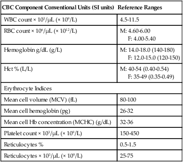
M, male; F, female; RBC, Red blood cell; SI, standard international; WBC, white blood cell.

CBC analyzers examine whole blood using various technologies and software to count cells and assess their volume and internal structures. However, CBC results are reviewed and verified by laboratory scientists before they are reported. The following discussion focuses first on WBCs and the differential count, and then shifts to RBCs and platelets. Coagulation studies are discussed last.

White Blood Cells

WBCs function as part of the immune system in protecting the body from various pathogenic microorganisms and foreign antigens. WBCs originate from hematopoietic stem cells in the bone marrow, develop into specific cell lineages through the influence of growth factors, and are released into the peripheral blood when mature. The WBCs that are normally present in the peripheral blood include segmented neutrophils, bands, eosinophils, basophils, lymphocytes, and monocytes.

White Blood Cell Differential Count

CBC analyzers determine the total number of WBCs per microliter or liter of blood and perform an automated differential count in which the percentage and absolute number of each type of WBC is determined. If the instrument count is abnormal, or a sample is flagged for suspected abnormal or immature cells, a manual slide review and differential count is performed by a laboratory scientist. In addition, the morphology of all the cells is assessed, and the presence of immature and abnormal cells is noted.

The relative count is the percentage of a particular cell among all the WBCs counted. The absolute count for that cell type is determined by multiplying its percentage or relative count by the total WBC count. CBC analyzers automatically calculate the absolute counts for each type of WBC. Table 7-2 provides sample reference ranges for the relative and absolute counts of each type of WBC.

TABLE 7-2

Sample Reference Ranges for the Differential White Blood Cell Count in Adults

Cell Type Relative Absolute
Segmented neutrophils 50-70 2.3-8.1
Bands 0-5 0-0.6
Eosinophils 1-3 0-0.4
Basophils 0-1 0-0.1
Lymphocytes 20-45 0.8-4.8
Monocytes 2-11 0.45-1.3

image

% × 103/μL (× 109/L)

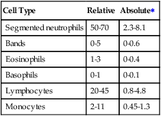
Because the relative count is influenced by increases or decreases of the other WBC types, the absolute count provides a more useful quantitative assessment. In a simplified example, if an adult patient had a total WBC count of 20 ×109/L with a relative count of 85% neutrophils and 15% lymphocytes, the absolute counts would be 17 × 109/L for neutrophils and 3 × 10⁹/L for lymphocytes. Comparing these results to the reference ranges in Table 7-2, there is both a relative and an absolute increase in neutrophils. On the other hand, although the relative count for the lymphocytes is decreased, the absolute count is well within the reference range.

White Blood Cell Functions

Neutrophils, eosinophils, and basophils are included in a general category of cells called granulocytes because of their prominent granules. However, they have different functions.

Segmented neutrophils (previously called polymorphonuclear leukocytes) are the most numerous cell type in the peripheral blood, constituting 50% to 70% of circulating WBCs. A band is a slightly less mature neutrophil in which the nucleus has not yet segmented. Bands normally represent only 0% to 5% of the circulating WBCs.

The primary function of neutrophils is to destroy invading microorganisms, foreign material, and dead cells. They accomplish this by phagocytosis, a process in which the neutrophil engulfs and destroys the particles through release of various enzymes and reactive molecules.

Neutrophils are produced and stored in the bone marrow, a process that takes 8 to 12 days. However, if the demand for neutrophils increases—as may occur in an acute bacterial infection— their time in the bone marrow may be shortened to as few as 2 days. In these cases, some immature neutrophils may be released. Once released into the peripheral blood, neutrophils have a very short half-life of 6 to 8 hours.

In the peripheral blood, neutrophils are continuously exchanged between two intravascular pools, with about half the cells in the circulating pool and half in the marginated pool. The circulating neutrophils are those counted in the CBC. Marginated neutrophils adhere to the walls of the blood vessels and are not counted. Neutrophils are able to rapidly shift from one pool to the other based on physiologic conditions (discussed subsequently).

Eosinophils are a type of granulocyte with large granules that stain bright orange or pink, whereas basophils have large granules that stain dark blue or purple. Eosinophils normally constitute 1% to 3% of WBCs, whereas basophils are even more rare at 0% to 1%. Both cell types are involved in immune system regulation, control of parasitic infections, and allergic reactions. Eosinophils also accumulate at the site of allergic reactions.

Lymphocytes constitute 20% to 45% of circulating WBCs in adults; in healthy children, the relative and absolute lymphocyte count is higher. Lymphocytes are particularly important in the body’s defense against foreign microorganisms and cells. There are three major types of lymphocytes: T cells, B cells, and NK (natural killer) cells. T and B cells participate in the body’s adaptive or specific immune response by recognizing foreign antigens and tagging them for destruction. T cells are involved in cell-mediated immunity, which is particularly important in eliminating viruses and other intracellular organisms. B cells are involved in humoral immunity and develop into antibody-producing cells. NK cells are able to destroy certain virally infected cells and tumor cells.

The different lymphocyte types appear similar on a peripheral blood smear. Enumeration of the different types of lymphocytes is not done routinely but can be accomplished using monoclonal antibodies. Approximately 85% of circulating lymphocytes are T cells, with B cells making up 10% to 15% and NK cells less than 2%. T lymphocytes also can be separated into subcategories. For example, knowledge of the CD4+ T-lymphocyte cell count provides important information about the severity and prognosis of patients with acquired immunodeficiency syndrome (AIDS; see later discussion of lymphocytopenia).

Monocytes are the largest WBCs normally seen in the peripheral blood and constitute 2% to 11% of circulating WBCs. In tissues, the monocyte is known as a macrophage. The primary functions of the monocyte are phagocytosis of organisms and other foreign material invading the body and initiation and regulation of the specific immune response with T lymphocytes. In the lung, alveolar macrophages play a key role in clearing inhaled particulate matter.

Nonmalignant White Blood Cell Abnormalities

Leukocytosis is an increase in the WBC count above the reference range, whereas leukopenia is a decrease in the WBC count below the reference range. Abnormalities in the WBC count can be defined more specifically by referring to the specific cell type that is increased or decreased using similar terminology. For example, an increase in neutrophils is called neutrophilia, and a decrease is called neutropenia. Increases in the cell counts may be primary (a result of uncontrolled proliferation of cells in the bone marrow) or secondary (a result of stimulation of the bone marrow secondary to other diseases or disorders). Similarly, decreases in cell counts may be caused by either primary bone marrow failure or increased destruction of the cells peripherally. Bone marrow failure can occur as a side effect of various drugs and disorders (secondary) or as a result of unknown causes (primary or idiopathic).

Neutrophilia is a common response to acute bacterial infections, such as bacterial pneumonia (Box 7-1). Neutrophilia also may occur in response to fungal, parasitic, or early viral infections. In addition, neutrophilia also occurs in inflammation due to autoimmune disorders or tissue damage that occurs after surgery, burns, myocardial infarction, or traumatic injury. Acute hemorrhage or hemolysis, metabolic disorders (such as acidosis or uremia), and certain drugs, toxins, and chemicals also cause neutrophilia. When the bone marrow is stimulated to release neutrophils at a rate faster than it can produce them, it releases them at increasingly more immature stages. This is called a left shift. The degree of the left shift and the severity of the neutrophilia usually correlate with the severity of the infection.

Pseudoneutrophilia, also called physiologic neutrophilia, occurs when marginated neutrophils are shifted to the circulating pool and are counted in the CBC. This type of neutrophilia is immediate and typically transient, lasting less than an hour. Because these are mature neutrophils, there is no spike in band-type cells as seen in pathologic neutrophilia.

Neutropenia may occur as a result of decreased bone marrow production or increased destruction of circulating neutrophils (Box 7-2). Many drugs, some chemicals, and radiation therapy can cause neutropenia by destroying the hematopoietic cells in the bone marrow, thus decreasing bone marrow production. One of the most common causes of neutropenia is cancer chemotherapy. Because of their short half-life, the neutrophils are the first blood cell type to decrease with chemotherapy. The absolute neutrophil count is closely monitored during chemotherapy, and adjustments in therapy are made if it drops too low. Other causes of decreased production include acquired and hereditary defects in hematopoietic stem cells, infiltration of cancer cells into the bone marrow, and deficiencies of vitamin B12 or folate. Neutrophils can also be quickly depleted by overwhelming infections or be destroyed as a result of the production of antibodies against them. Pseudoneutropenia is a transient shift of the neutrophils in the circulating pool to the marginated pool. It may be caused by bacterial endotoxins or hypersensitivity reactions.

Neutrophils are the first-line defense against microorganisms, so patients with neutropenia have a high risk for developing life-threatening bacterial or fungal infections. The lower the absolute neutrophil count and the longer the duration of the neutropenia, the greater the risk for serious infection.

Eosinophilia (increase in eosinophils) is often seen in parasitic infestations and allergic states (such as hay fever, dermatitis, and drug reactions). Patients with extrinsic or atopic asthma often have eosinophilia. Basophilia (increase in basophils) is usually associated with myeloproliferative neoplasms (discussed later in the chapter).

Lymphocytosis (increase in lymphocytes) is typically seen in viral infections and certain bacterial (pertussis) and parasitic (toxoplasmosis) infections. In some viral infections, especially infectious mononucleosis, many of the lymphocytes are enlarged and have a characteristic appearance, being labeled as reactive or variant lymphocytes. Interpretation of lymphocytosis should take into account the patient’s age because children normally have higher counts than adults. Lymphocytopenia (decrease in lymphocytes) is seen in acquired and congenital immune deficiency states and in various conditions such as acute inflammation, malnutrition, and after treatment with chemotherapy, radiation, or corticosteroids. Lymphocytopenia is an important feature of human immunodeficiency virus (HIV) infection, the virus that causes AIDS. The virus infects and destroys the CD4+ helper T lymphocytes, resulting in their depletion. As the CD4+ cells decline, the immune system becomes more compromised, leading to increased risk for certain cancers and infections. Peripheral blood CD4+ counts, along with the HIV viral load, are used to monitor the progression of the disease.

Monocytosis (increase in monocytes) is characteristic of certain infections, including tuberculosis, syphilis, typhoid fever, and subacute bacterial endocarditis. A monocytosis often signals the recovery stage of an acute infection. Monocytosis also may occur in inflammatory conditions and autoimmune states.

Malignant White Blood Cell Abnormalities

Leukemias, myeloproliferative neoplasms, and myelodysplastic syndromes are the primary hematologic malignancies involving the bone marrow and peripheral blood. Leukemias result from the uncontrolled proliferation (growth) of a specific type of WBC and may be either acute or chronic. Myeloproliferative neoplasms encompass a spectrum of diseases resulting from an abnormality in stem cells (the precursor of all blood cells). Myeloproliferative neoplasms may have a variable blood picture as the disease progresses, and they sometimes terminate as an acute leukemia. Myelodysplastic syndromes are characterized by decreases in WBCs, RBCs, and platelets in the peripheral blood due to a stem cell defect.

In acute leukemia, there is a mutation that causes a maturation arrest in a blood cell precursor at an early stage of development. Typically, blasts, which are the most immature stage of a cell type, accumulate in the bone marrow and peripheral blood. These blasts quickly replace all other cells in the bone marrow and, if left untreated, will cause a rapid death of the patient. WBC counts are variable, but the key feature in the WBC differential is the presence of blasts. The normal WBCs, along with RBCs and platelets, are decreased. Acute lymphoblastic leukemia usually occurs in young children, whereas acute myeloid leukemia most often occurs in infants and older adults. Acute leukemias are classified by the World Health Organization according to the presence of recurrent genetic abnormalities in the leukemia cells.

Chronic leukemias result from a slower proliferation and accumulation of more mature cells in the peripheral blood and bone marrow. Chronic lymphocytic leukemia is the most common type of leukemia and occurs predominantly in elderly people. The WBC count is increased, and the differential shows a preponderance of mature lymphocytes. Chronic myelogenous leukemia (CML) is a myeloproliferative neoplasm that occurs predominantly in middle-aged adults. It is caused by a mutation in a stem cell that results in uncontrolled proliferation of granulocytes. The WBC count is increased, and the bone marrow and peripheral blood contain vast numbers of neutrophils, with immature neutrophils in all stages of maturation, as well as eosinophils and basophils. Platelets are also increased, and there is progressive anemia. Without treatment, CML terminates in acute leukemia, known as blast transformation.

Other myeloproliferative neoplasms include polycythemia vera, primary myelofibrosis, and essential thrombocythemia. Polycythemia vera is characterized by a proliferation of granulocytic, erythrocytic, and platelet precursors in the bone marrow with an increase in WBCs, RBCs, and platelets in the peripheral blood. Myelofibrosis is characterized by defective hematopoiesis caused by the excessive growth of fibrous tissue (fibrosis) in the bone marrow. Essential thrombocythemia primarily involves the excessive proliferation of megakaryocytes, with increased platelets in the peripheral blood. Myelodysplastic syndromes result from stem cell mutations that cause ineffective blood cell production in the bone marrow and a progressive decrease in WBCs, RBCs, and platelets in the peripheral blood. They occur predominantly in the elderly.

Red Blood Cells

RBCs are produced in the bone marrow by maturation of nucleated erythrocytic precursor cells known as normoblasts. Under normal circumstances, as the normoblasts mature, the nuclei become smaller and more condensed, and the cytoplasm acquires a pink color as a result of the development of hemoglobin. Before the RBC is released from the bone marrow to circulate in the peripheral blood, the nucleus is removed. RBCs have a life span of approximately 120 days.

Normal RBCs assume the shape of a biconcave disk to facilitate their primary function of carrying oxygen and to provide maximal deformability to bend as they pass through small capillaries. The major component of mature RBCs (erythrocytes) is hemoglobin, which imparts to blood its normal red color when carrying oxygen. The RBC count is reported in number of cells per microliter or liter of blood (see Table 7-1 for reference ranges). An adequate number of RBCs with an adequate concentration of hemoglobin and a functionally normal hemoglobin molecule are needed for transport of sufficient oxygen from the lungs to the tissues.
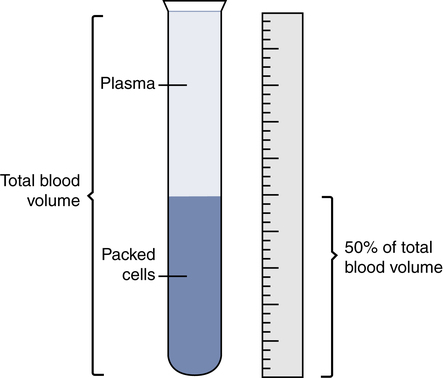
Red Blood Cell Indices

Three erythrocyte indices are the mean cell volume (MCV), mean cell hemoglobin (MCH), and mean cell hemoglobin concentration (MCHC). They are calculated from the RBC count, hemoglobin, and Hct. The MCV is the average volume of the patient’s RBCs. A decrease in the MCV indicates the RBCs are smaller, or microcytic, whereas an increase in the MCV indicates the RBCs are larger, or macrocytic. A normal MCV indicates the RBCs are normal in volume, or normocytic, although the presence of an equal number of smaller and larger RBCs could produce a normal MCV. The MCH is the average weight of hemoglobin in the patient’s RBCs. The interpretation of the MCH correlates with that of the MCV. The MCHC is the concentration of hemoglobin in the patient’s RBCs. A decrease in the MCHC indicates the RBCs have less hemoglobin, called hypochromic, whereas a normal MCHC indicates the RBCs have a normal complement of hemoglobin, called normochromic.

Red Blood Cell Abnormalities

Anemia is a decrease in the oxygen carrying capacity of the blood due to a quantitative deficiency or a functional defect in hemoglobin. Although the RBC count and Hct also decrease, anemia is clinically defined by a decrease in the hemoglobin concentration below the reference range for an individual’s gender and age.

One of the body’s responses to hypoxia is an increase in secretion of erythropoietin by the kidneys, which accelerates the maturation and release of new RBCs into the circulation. The circulatory system also responds to anemia by increasing the heart rate, respiration rate, and cardiac output to deliver oxygen more quickly to the tissues.

General symptoms of anemia include fatigue and dyspnea, but various other symptoms may also occur depending on the cause of the anemia. In severe anemia, tachycardia and hypotension also occur, which if untreated can lead to cardiac failure. The severity of the symptoms depends on the degree of hemoglobin reduction, the state of the patient’s cardiovascular system, and the rate of development and duration of the anemia.

Anemias may be classified by pathophysiology into those caused by a decrease in RBC production and those caused by an increase in RBC destruction or loss. However, a more practical approach is to use the MCV to classify anemias into microcytic, macrocytic, or normocytic types.

The most common type of anemia worldwide is the result of iron deficiency. Iron deficiency anemia occurs when there is a dietary deficiency of iron, increased need for iron that is not met (pregnancy, infancy, adolescent growth spurts), malabsorption of iron, or chronic blood loss. In chronic blood loss, the iron contained in the RBCs slowly leaves the body instead of being recycled. Iron is required for hemoglobin synthesis, and with sustained, low iron levels, the bone marrow begins to produce smaller RBCs with a decreased amount of hemoglobin, and the RBC indices become microcytic and hypochromic.

Anemia also can result from chronic inflammation that causes an impairment of iron regulation and RBC production in the bone marrow. The resulting anemia is often normocytic, but it can be microcytic in diseases of long duration. Macrocytic anemia is most often caused by either folate or vitamin B12 deficiency. Low folate or vitamin B12 levels impair DNA synthesis in the nucleus and result in ineffective blood cell production and macrocytic RBCs.

The hemolytic anemias constitute a large group of disorders in which excessive destruction of RBCs in the circulation exceeds the capacity of the bone marrow to replace the cells that are lost. Hemolytic anemia may be due to intrinsic defects in the RBCs (such as sickle cell anemia) or abnormalities extrinsic to the RBCs (such as hemolysis caused by antibodies, malaria, or drugs).

Hemoglobin may be converted to an inactive form through oxidation, denaturation, or other chemical reaction. The most common forms of inactive hemoglobins are carboxyhemoglobin (COHb) and methemoglobin (metHb). The measurement of these abnormal Hb combinations and their impact on oxygen transport are discussed in detail in Chapter 8.

Polycythemia is an increase in the RBC count, hemoglobin, and Hct and may be primary, secondary, or relative (spurious). Primary polycythemia is uncommon and is caused by an uncontrolled proliferation of hematopoietic cells within the bone marrow (polycythemia vera, previously discussed)

Secondary polycythemia is more common and is seen in patients who have chronic stimulation of the bone marrow to produce more RBCs secondary to some other disorder or condition. Examples include patients with chronic hypoxemia due to disease (e.g., chronic obstructive pulmonary disease [COPD]), obstructive sleep apnea, pulmonary fibrosis, chronic heart failure) or as compensation for the low oxygen pressures breathed by those living at high altitude. In these cases, chronic hypoxemia stimulates renal production of erythropoietin, causing the bone marrow to produce and release additional RBCs to compensate for the oxygen deficit.

Heavy smokers also may exhibit secondary polycythemia. The carbon monoxide associated with cigarette smoke binds tightly with the hemoglobin and reduces oxygen transport. This results in a functional anemia to which the bone marrow responds by increasing RBC production.

In the presence of significant hypoxia due to severe anemia, the maturation time of RBCs in the bone marrow is decreased, resulting in the release of immature nucleated RBCs (NRBCs), which are not normally seen in the blood of adults. As a result of the hypoxic conditions in utero, NRBCs are present in fetal blood and newborns but disappear 3 to 4 days after birth. NRBCs also occur in patients with some myeloproliferative neoplasms.

Both primary and secondary polycythemias involve an absolute increase in the total RBC mass. Sometimes, relative polycythemia occurs because of a decrease in the plasma volume. This is seen in patients who are dehydrated. Relative polycythemia does not represent a true increase in the number of circulating RBCs.

Although polycythemia is helpful in increasing the oxygen-carrying capacity of the blood, it can be detrimental to the heart and circulatory system. Polycythemia increases the viscosity of the blood, causing both an increased cardiac workload and a greater risk for clot formation and thrombosis. Treatment focuses on management of the underlying cause and in some patients may include restoring a normal Hct by controlled removal of whole blood through phlebotomy.

Reticulocyte Count

The reticulocyte is the final erythrocyte development stage before the RBC is fully mature. These slightly immature RBCs lack a nucleus and differ from mature RBCs in that they are slightly larger and have not yet assumed a biconcave shape. A reticulocyte count is performed by obtaining the percentage of reticulocytes among the RBCs. Normally about 1% of the circulating RBCs are reticulocytes, with slightly higher values in newborns. The reticulocyte count is also reported as an absolute count by multiplying the reticulocyte percentage by the RBC count. Reference ranges are provided in Table 7-1.

The reticulocyte count helps to assess the bone marrow response to an anemia. If the absolute reticulocyte count is high in a patient with anemia, it indicates that the bone marrow is responding to the anemia by increasing production of RBCs. In this case the cause of the anemia is likely due to excessive peripheral blood loss or destruction. However, if the reticulocyte count is low, then the anemia is likely the result of decreased bone marrow production.

Platelet Count

Platelets are the smallest cells in the peripheral blood. After injury to a blood vessel, they participate in clot formation at the site of the injury to stop the bleeding. The platelet count routinely is provided as part of the CBC. Table 7-1 provides the reference range for the platelet count. Like other blood cells, platelets may be decreased as a result of increased destruction in the peripheral blood or decreased production in the bone marrow. A reduction in the platelet count below the reference range is called thrombocytopenia.

Thrombocytopenia usually manifests as small skin hemorrhages (petechiae and ecchymoses) and bleeding from mucosal surfaces (such as epistaxis or menorrhagia). When the platelet count is decreased significantly (<20 × 103/µL),patients are more likely to have bleeding problems, especially with trauma such as surgery or arterial punctures. However, when the platelet count becomes extremely low (<10 × 103/µL), the patient is at risk for serious spontaneous bleeding, including intracranial hemorrhage. There are many disorders that cause thrombocytopenia, including side effects of drugs such as chemotherapy or heparin, bone marrow diseases, and immune thrombocytopenic purpura, an autoimmune disorder in which autoantibodies are produced against the platelets, marking them for destruction in the spleen.

An increase in platelets (thrombocytosis